6P2U

Structure of Mortalin-NBD with N6-propargyladenosine-5'-diphosphate

  • Classification: CHAPERONE
  • Organism(s): Homo sapiens
  • Expression System: Escherichia coli BL21(DE3)
  • Mutation(s): No 

  • Deposited: 2019-05-22 Released: 2019-06-19 
  • Deposition Author(s): Moseng, M.A., Page, R.C.
  • Funding Organization(s): National Institutes of Health/National Institute of General Medical Sciences (NIH/NIGMS), National Science Foundation (NSF, United States)

Experimental Data Snapshot

  • Method: X-RAY DIFFRACTION
  • Resolution: 2.00 Å
  • R-Value Free: 0.238 
  • R-Value Work: 0.215 
  • R-Value Observed: 0.217 

wwPDB Validation   3D Report Full Report


Ligand Structure Quality Assessment 


This is version 1.4 of the entry. See complete history


Literature

2- and N6-functionalized adenosine-5'-diphosphate analogs for the inhibition of mortalin.

Moseng, M.A.Nix, J.C.Page, R.C.

(2019) FEBS Lett 593: 2030-2039

  • DOI: https://doi.org/10.1002/1873-3468.13475
  • Primary Citation of Related Structures:  
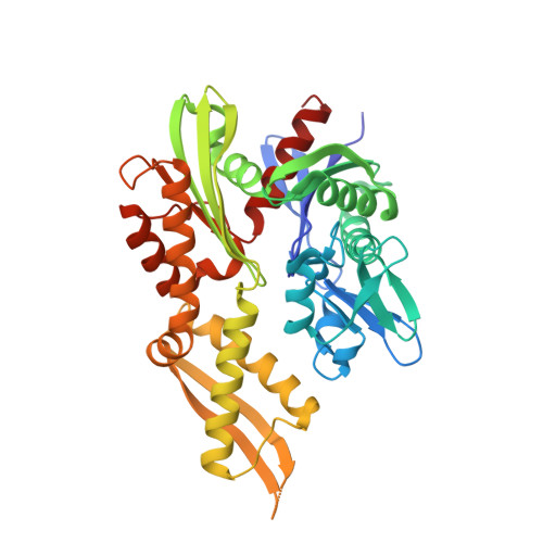
    6P2U

  • PubMed Abstract: 

    Our early efforts to find a covalent inhibitor of mortalin, a member of the 70 kD heat shock protein (Hsp70) family, led us to solve the structure of the mortalin nucleotide-binding domain (NBD) in complex with N6-propargyladenosine-5'-diphosphate. The acquired structure emphasizes the ability of the nucleotide-binding pocket to accommodate modified ADP compounds. A library of ADP analogs modified at either the 2- or N6-positions of adenosine was screened against the mortalin-NBD. Competitive inhibition and binding assays of the analogs demonstrate that modifications at the 2- or N6-positions have potential to bind and inhibit mortalin uniquely compared to other Hsp70 homologs, and that modifications at the 2-position confer the greatest selectivity in binding and inhibition of the mortalin-NBD.


  • Organizational Affiliation

    Department of Chemistry and Biochemistry, Miami University, Oxford, OH, USA.


Macromolecules
Find similar proteins by:  (by identity cutoff)  |  3D Structure
Entity ID: 1
MoleculeChains Sequence LengthOrganismDetailsImage
Stress-70 protein, mitochondrial385Homo sapiensMutation(s): 0 
Gene Names: HSPA9GRP75HSPA9Bmt-HSP70
UniProt & NIH Common Fund Data Resources
Find proteins for P38646 (Homo sapiens)
Explore P38646 
Go to UniProtKB:  P38646
PHAROS:  P38646
GTEx:  ENSG00000113013 
Entity Groups  
Sequence Clusters30% Identity50% Identity70% Identity90% Identity95% Identity100% Identity
UniProt GroupP38646
Sequence Annotations
Expand
  • Reference Sequence
Small Molecules
Ligands 3 Unique
IDChains Name / Formula / InChI Key2D Diagram3D Interactions
NO7 (Subject of Investigation/LOI)
Query on NO7

Download Ideal Coordinates CCD File 
B [auth A]9-{5-O-[(S)-hydroxy(phosphonooxy)phosphoryl]-alpha-D-ribofuranosyl}-N-(prop-2-yn-1-yl)-9H-purin-6-amine
C13 H17 N5 O10 P2
DYWGWOPDXRINLT-HMMKDHSLSA-N
PO4
Query on PO4

Download Ideal Coordinates CCD File 
C [auth A]PHOSPHATE ION
O4 P
NBIIXXVUZAFLBC-UHFFFAOYSA-K
MG
Query on MG

Download Ideal Coordinates CCD File 
D [auth A]MAGNESIUM ION
Mg
JLVVSXFLKOJNIY-UHFFFAOYSA-N
Experimental Data & Validation

Experimental Data

  • Method: X-RAY DIFFRACTION
  • Resolution: 2.00 Å
  • R-Value Free: 0.238 
  • R-Value Work: 0.215 
  • R-Value Observed: 0.217 
  • Space Group: P 21 21 21
Unit Cell:
Length ( Å )Angle ( ˚ )
a = 51.04α = 90
b = 68.25β = 90
c = 121.22γ = 90
Software Package:
Software NamePurpose
PHENIXrefinement
XDSdata reduction
XDSdata scaling
PHASERphasing

Structure Validation

View Full Validation Report


Ligand Structure Quality Assessment 


Entry History & Funding Information

Deposition Data


Funding OrganizationLocationGrant Number
National Institutes of Health/National Institute of General Medical Sciences (NIH/NIGMS)United StatesGM128595
National Science Foundation (NSF, United States)United StatesMCB 15-52113

Revision History  (Full details and data files)

  • Version 1.0: 2019-06-19
    Type: Initial release
  • Version 1.1: 2019-07-03
    Changes: Data collection, Database references
  • Version 1.2: 2019-08-28
    Changes: Data collection, Database references
  • Version 1.3: 2019-11-27
    Changes: Author supporting evidence
  • Version 1.4: 2023-10-11
    Changes: Data collection, Database references, Refinement description