6UOK

Y271G DNA polymerase beta substrate complex with templating cytosine and incoming r8-oxo-GTP


Experimental Data Snapshot

  • Method: X-RAY DIFFRACTION
  • Resolution: 2.55 Å
  • R-Value Free: 0.279 
  • R-Value Work: 0.224 
  • R-Value Observed: 0.229 

wwPDB Validation   3D Report Full Report


Ligand Structure Quality Assessment 


This is version 1.3 of the entry. See complete history


Literature

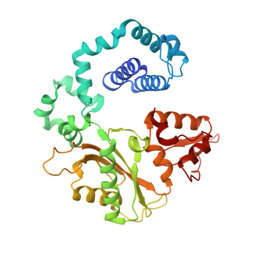
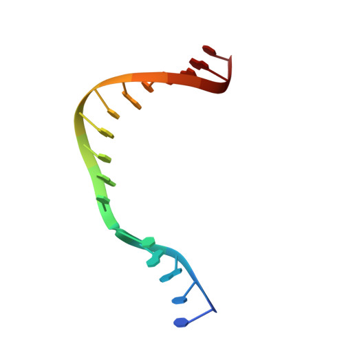
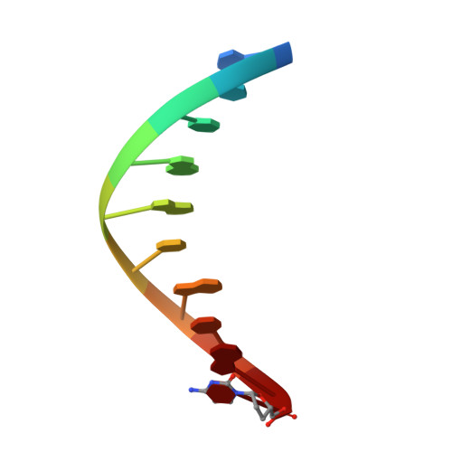
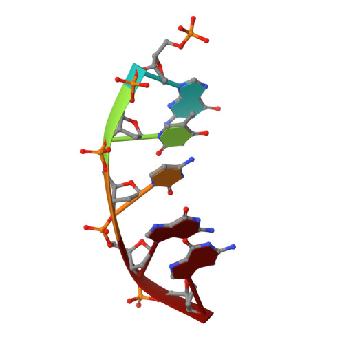
Molecular and structural characterization of oxidized ribonucleotide insertion into DNA by human DNA polymerase beta.

Smith, M.R.Alnajjar, K.S.Hoitsma, N.M.Sweasy, J.B.Freudenthal, B.D.

(2020) J Biol Chem 295: 1613-1622

  • DOI: https://doi.org/10.1074/jbc.RA119.011569
  • Primary Citation of Related Structures:  
    6UOK, 6UOL, 6UOM

  • PubMed Abstract: 

    During oxidative stress, inflammation, or environmental exposure, ribo- and deoxyribonucleotides are oxidatively modified. 8-Oxo-7,8-dihydro-2'-guanosine (8-oxo-G) is a common oxidized nucleobase whose deoxyribonucleotide form, 8-oxo-dGTP, has been widely studied and demonstrated to be a mutagenic substrate for DNA polymerases. Guanine ribonucleotides are analogously oxidized to r8-oxo-GTP, which can constitute up to 5% of the rGTP pool. Because ribonucleotides are commonly misinserted into DNA, and 8-oxo-G causes replication errors, we were motivated to investigate how the oxidized ribonucleotide is utilized by DNA polymerases. To do this, here we employed human DNA polymerase β (pol β) and characterized r8-oxo-GTP insertion with DNA substrates containing either a templating cytosine (nonmutagenic) or adenine (mutagenic). Our results show that pol β has a diminished catalytic efficiency for r8-oxo-GTP compared with canonical deoxyribonucleotides but that r8-oxo-GTP is inserted mutagenically at a rate similar to those of other common DNA replication errors ( i.e. ribonucleotide and mismatch insertions). Using FRET assays to monitor conformational changes of pol β with r8-oxo-GTP, we demonstrate impaired pol β closure that correlates with a reduced insertion efficiency. X-ray crystallographic analyses revealed that, similar to 8-oxo-dGTP, r8-oxo-GTP adopts an anti conformation opposite a templating cytosine and a syn conformation opposite adenine. However, unlike 8-oxo-dGTP, r8-oxo-GTP did not form a planar base pair with either templating base. These results suggest that r8-oxo-GTP is a potential mutagenic substrate for DNA polymerases and provide structural insights into how r8-oxo-GTP is processed by DNA polymerases.


  • Organizational Affiliation

    Department of Biochemistry and Molecular Biology, University of Kansas Medical Center, Kansas City, Kansas 66160; Department of Cancer Biology, University of Kansas Medical Center, Kansas City, Kansas 66160.


Macromolecules

Find similar proteins by:  (by identity cutoff)  |  3D Structure
Entity ID: 4
MoleculeChains Sequence LengthOrganismDetailsImage
DNA polymerase betaD [auth F],
E [auth A]
335Homo sapiensMutation(s): 1 
Gene Names: POLB
EC: 2.7.7.7 (PDB Primary Data), 4.2.99 (PDB Primary Data)
UniProt & NIH Common Fund Data Resources
Find proteins for P06746 (Homo sapiens)
Explore P06746 
Go to UniProtKB:  P06746
PHAROS:  P06746
GTEx:  ENSG00000070501 
Entity Groups  
Sequence Clusters30% Identity50% Identity70% Identity90% Identity95% Identity100% Identity
UniProt GroupP06746
Sequence Annotations
Expand
  • Reference Sequence

Find similar nucleic acids by:  Sequence   |   3D Structure  

Entity ID: 1
MoleculeChains LengthOrganismImage
DNA (5'-D(*CP*CP*GP*AP*CP*CP*GP*CP*GP*CP*AP*TP*CP*AP*GP*C)-3')A [auth B],
F [auth T]
16synthetic construct
Sequence Annotations
Expand
  • Reference Sequence

Find similar nucleic acids by:  Sequence   |   3D Structure  

Entity ID: 2
MoleculeChains LengthOrganismImage
DNA (5'-D(*GP*CP*TP*GP*AP*TP*GP*CP*GP*(DOC))-3')B [auth C],
G [auth P]
10synthetic construct
Sequence Annotations
Expand
  • Reference Sequence

Find similar nucleic acids by:  Sequence   |   3D Structure  

Entity ID: 3
MoleculeChains LengthOrganismImage
DNA (5'-D(P*GP*TP*CP*GP*G)-3')C [auth E],
H [auth D]
5synthetic construct
Sequence Annotations
Expand
  • Reference Sequence
Small Molecules
Ligands 6 Unique
IDChains Name / Formula / InChI Key2D Diagram3D Interactions
8GT (Subject of Investigation/LOI)
Query on 8GT

Download Ideal Coordinates CCD File 
P [auth F],
W [auth A]
8-OXO-GUANOSINE-5'-TRIPHOSPHATE
C10 H16 N5 O15 P3
JCHLKIQZUXYLPW-UMMCILCDSA-N
EDO
Query on EDO

Download Ideal Coordinates CCD File 
S [auth A]1,2-ETHANEDIOL
C2 H6 O2
LYCAIKOWRPUZTN-UHFFFAOYSA-N
MN
Query on MN

Download Ideal Coordinates CCD File 
L [auth F]
M [auth F]
N [auth F]
Q [auth F]
R [auth F]
L [auth F],
M [auth F],
N [auth F],
Q [auth F],
R [auth F],
V [auth A]
MANGANESE (II) ION
Mn
WAEMQWOKJMHJLA-UHFFFAOYSA-N
CL
Query on CL

Download Ideal Coordinates CCD File 
I [auth B]CHLORIDE ION
Cl
VEXZGXHMUGYJMC-UHFFFAOYSA-M
MG
Query on MG

Download Ideal Coordinates CCD File 
Y [auth D]MAGNESIUM ION
Mg
JLVVSXFLKOJNIY-UHFFFAOYSA-N
NA
Query on NA

Download Ideal Coordinates CCD File 
J [auth E]
K [auth F]
O [auth F]
T [auth A]
U [auth A]
J [auth E],
K [auth F],
O [auth F],
T [auth A],
U [auth A],
X [auth A]
SODIUM ION
Na
FKNQFGJONOIPTF-UHFFFAOYSA-N
Experimental Data & Validation

Experimental Data

  • Method: X-RAY DIFFRACTION
  • Resolution: 2.55 Å
  • R-Value Free: 0.279 
  • R-Value Work: 0.224 
  • R-Value Observed: 0.229 
  • Space Group: P 1 21 1
Unit Cell:
Length ( Å )Angle ( ˚ )
a = 54.687α = 90
b = 79.957β = 97.642
c = 100.832γ = 90
Software Package:
Software NamePurpose
PHENIXrefinement
HKL-3000data collection
HKL-3000data scaling
PDB_EXTRACTdata extraction
HKL-3000data reduction
PHENIXphasing

Structure Validation

View Full Validation Report


Ligand Structure Quality Assessment 


Entry History & Funding Information

Deposition Data


Funding OrganizationLocationGrant Number
National Institutes of Health/National Institute of Environmental Health Sciences (NIH/NIEHS)United States--

Revision History  (Full details and data files)

  • Version 1.0: 2020-01-08
    Type: Initial release
  • Version 1.1: 2020-01-15
    Changes: Database references
  • Version 1.2: 2020-02-19
    Changes: Database references
  • Version 1.3: 2023-10-11
    Changes: Data collection, Database references, Derived calculations, Refinement description