1B59

COMPLEX OF HUMAN METHIONINE AMINOPEPTIDASE-2 COMPLEXED WITH OVALICIN


Experimental Data Snapshot

  • Method: X-RAY DIFFRACTION
  • Resolution: 1.80 Å
  • R-Value Free: 0.222 
  • R-Value Work: 0.178 
  • R-Value Observed: 0.178 

wwPDB Validation   3D Report Full Report


Ligand Structure Quality Assessment 


This is version 1.3 of the entry. See complete history


Literature

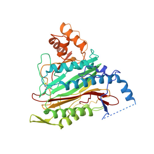
Structure of human methionine aminopeptidase-2 complexed with fumagillin.

Liu, S.Widom, J.Kemp, C.W.Crews, C.M.Clardy, J.

(1998) Science 282: 1324-1327

  • DOI: https://doi.org/10.1126/science.282.5392.1324
  • Primary Citation of Related Structures:  
    1B59, 1B6A, 1BN5, 1BOA

  • PubMed Abstract: 

    The fungal metabolite fumagillin suppresses the formation of new blood vessels, and a fumagillin analog is currently in clinical trials as an anticancer agent. The molecular target of fumagillin is methionine aminopeptidase-2 (MetAP-2). A 1.8 A resolution crystal structure of free and inhibited human MetAP-2 shows a covalent bond formed between a reactive epoxide of fumagillin and histidine-231 in the active site of MetAP-2. Extensive hydrophobic and water-mediated polar interactions with other parts of fumagillin provide additional affinity. Fumagillin-based drugs inhibit MetAP-2 but not MetAP-1, and the three-dimensional structure also indicates the likely determinants of this specificity. The structural basis for fumagillin's potency and specificity forms the starting point for structure-based drug design.


  • Organizational Affiliation

    J. Clardy, Department of Chemistry and Chemical Biology, Baker Laboratory, Cornell University, Ithaca, NY 14853-1301, USA.


Macromolecules
Find similar proteins by:  (by identity cutoff)  |  3D Structure
Entity ID: 1
MoleculeChains Sequence LengthOrganismDetailsImage
PROTEIN (METHIONINE AMINOPEPTIDASE)370Homo sapiensMutation(s): 0 
EC: 3.4.11.18
UniProt & NIH Common Fund Data Resources
Find proteins for P50579 (Homo sapiens)
Explore P50579 
Go to UniProtKB:  P50579
PHAROS:  P50579
GTEx:  ENSG00000111142 
Entity Groups  
Sequence Clusters30% Identity50% Identity70% Identity90% Identity95% Identity100% Identity
UniProt GroupP50579
Sequence Annotations
Expand
  • Reference Sequence
Small Molecules
Ligands 2 Unique
IDChains Name / Formula / InChI Key2D Diagram3D Interactions
OVA
Query on OVA

Download Ideal Coordinates CCD File 
D [auth A]3,4-DIHYDROXY-2-METHOXY-4-METHYL-3-[2-METHYL-3-(3-METHYL-BUT-2-ENYL) -OXIRANYL]-CYCLOHEXANONE
C16 H26 O5
UOXVFQCRPDLSFN-DGXTUMSLSA-N
CO
Query on CO

Download Ideal Coordinates CCD File 
B [auth A],
C [auth A]
COBALT (II) ION
Co
XLJKHNWPARRRJB-UHFFFAOYSA-N
Experimental Data & Validation

Experimental Data

  • Method: X-RAY DIFFRACTION
  • Resolution: 1.80 Å
  • R-Value Free: 0.222 
  • R-Value Work: 0.178 
  • R-Value Observed: 0.178 
  • Space Group: C 2 2 21
Unit Cell:
Length ( Å )Angle ( ˚ )
a = 89.723α = 90
b = 99.108β = 90
c = 101.292γ = 90
Software Package:
Software NamePurpose
MOSFLMdata reduction
SCALAdata scaling
CNSrefinement
CCP4data scaling

Structure Validation

View Full Validation Report


Ligand Structure Quality Assessment 


Entry History 

Deposition Data

Revision History  (Full details and data files)

  • Version 1.0: 2000-01-14
    Type: Initial release
  • Version 1.1: 2007-10-16
    Changes: Version format compliance
  • Version 1.2: 2011-07-13
    Changes: Version format compliance
  • Version 1.3: 2023-08-09
    Changes: Data collection, Database references, Derived calculations, Refinement description