1BO4

CRYSTAL STRUCTURE OF A GCN5-RELATED N-ACETYLTRANSFERASE: SERRATIA MARESCENS AMINOGLYCOSIDE 3-N-ACETYLTRANSFERASE


Experimental Data Snapshot

  • Method: X-RAY DIFFRACTION
  • Resolution: 2.30 Å
  • R-Value Free: 0.250 
  • R-Value Work: 0.201 
  • R-Value Observed: 0.201 

wwPDB Validation   3D Report Full Report


This is version 1.4 of the entry. See complete history


Literature

Crystal structure of a GCN5-related N-acetyltransferase: Serratia marcescens aminoglycoside 3-N-acetyltransferase.

Wolf, E.Vassilev, A.Makino, Y.Sali, A.Nakatani, Y.Burley, S.K.

(1998) Cell 94: 439-449

  • DOI: https://doi.org/10.1016/s0092-8674(00)81585-8
  • Primary Citation of Related Structures:  
    1BO4

  • PubMed Abstract: 

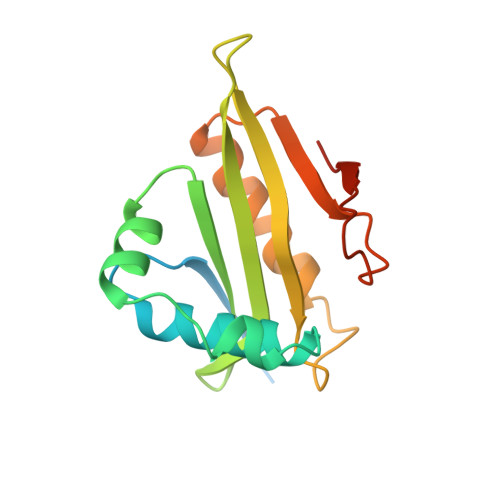
    The X-ray structure of a canonical GCN5-related N-acetyltransferase (GNAT), Serratia marcescens aminoglycoside 3-N-acetyltransferase, bound to coenzyme A (CoA) has been determined at 2.3 A resolution. The single domain alpha/beta protein resembles a cupped right hand wrapped around a cylinder and consists of a highly curved, six-stranded beta sheet of mixed polarity that is sandwiched between four alpha helices. The structure includes all four conserved GNAT motifs (C, D, A, and B) and represents the catalytic core of this large enzyme superfamily. Acetyl CoA recognition is mediated by a betaalpha structure derived from GNAT motif A, which presents an invariant Arg/Gln-X-X-Gly-X-Gly/Ala segment for hydrogen bonding with the cofactor. Motif B contributes acidic residues to the binding site for the positively charged antibiotic substrate.


  • Organizational Affiliation

    Laboratories of Molecular Biophysics, The Rockefeller University, New York, New York 10021, USA.


Macromolecules
Find similar proteins by:  (by identity cutoff)  |  3D Structure
Entity ID: 1
MoleculeChains Sequence LengthOrganismDetailsImage
PROTEIN (SERRATIA MARCESCENS AMINOGLYCOSIDE-3-N-ACETYLTRANSFERASE)
A, B
168Serratia marcescensMutation(s): 0 
Gene Names: AACC1
UniProt
Find proteins for Q53396 (Serratia marcescens)
Explore Q53396 
Go to UniProtKB:  Q53396
Entity Groups  
Sequence Clusters30% Identity50% Identity70% Identity90% Identity95% Identity100% Identity
UniProt GroupQ53396
Sequence Annotations
Expand
  • Reference Sequence
Small Molecules
Ligands 2 Unique
IDChains Name / Formula / InChI Key2D Diagram3D Interactions
COA
Query on COA

Download Ideal Coordinates CCD File 
D [auth A],
E [auth B]
COENZYME A
C21 H36 N7 O16 P3 S
RGJOEKWQDUBAIZ-IBOSZNHHSA-N
SPD
Query on SPD

Download Ideal Coordinates CCD File 
C [auth A]SPERMIDINE
C7 H19 N3
ATHGHQPFGPMSJY-UHFFFAOYSA-N
Experimental Data & Validation

Experimental Data

  • Method: X-RAY DIFFRACTION
  • Resolution: 2.30 Å
  • R-Value Free: 0.250 
  • R-Value Work: 0.201 
  • R-Value Observed: 0.201 
  • Space Group: C 1 2 1
Unit Cell:
Length ( Å )Angle ( ˚ )
a = 78.54α = 90
b = 102.26β = 93.73
c = 50.14γ = 90
Software Package:
Software NamePurpose
SHARPphasing
CNSrefinement
MOSFLMdata reduction
CCP4data scaling

Structure Validation

View Full Validation Report


Entry History 

Deposition Data

Revision History  (Full details and data files)

  • Version 1.0: 1998-10-07
    Type: Initial release
  • Version 1.1: 2007-10-16
    Changes: Version format compliance
  • Version 1.2: 2011-07-13
    Changes: Derived calculations, Version format compliance
  • Version 1.3: 2021-02-03
    Changes: Database references, Derived calculations, Structure summary
  • Version 1.4: 2024-02-07
    Changes: Data collection, Database references