1C0K

CRYSTAL STRUCTURE ANALYSIS OF D-AMINO ACID OXIDASE IN COMPLEX WITH L-LACTATE


Experimental Data Snapshot

  • Method: X-RAY DIFFRACTION
  • Resolution: 1.46 Å

wwPDB Validation   3D Report Full Report


This is version 2.0 of the entry. See complete history


Literature

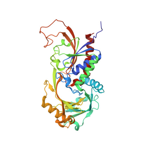
The x-ray structure of D-amino acid oxidase at very high resolution identifies the chemical mechanism of flavin-dependent substrate dehydrogenation.

Umhau, S.Pollegioni, L.Molla, G.Diederichs, K.Welte, W.Pilone, M.S.Ghisla, S.

(2000) Proc Natl Acad Sci U S A 97: 12463-12468

  • DOI: https://doi.org/10.1073/pnas.97.23.12463
  • Primary Citation of Related Structures:  
    1C0K, 1C0L, 1C0P

  • PubMed Abstract: 

    Flavin is one of the most versatile redox cofactors in nature and is used by many enzymes to perform a multitude of chemical reactions. d-Amino acid oxidase (DAAO), a member of the flavoprotein oxidase family, is regarded as a key enzyme for the understanding of the mechanism underlying flavin catalysis. The very high-resolution structures of yeast DAAO complexed with d-alanine, d-trifluoroalanine, and l-lactate (1.20, 1.47, and 1.72 A) provide strong evidence for hydride transfer as the mechanism of dehydrogenation. This is inconsistent with the alternative carbanion mechanism originally favored for this type of enzymatic reaction. The step of hydride transfer can proceed without involvement of amino acid functional groups. These structures, together with results from site-directed mutagenesis, point to orbital orientation/steering as the major factor in catalysis. A diatomic species, proposed to be a peroxide, is found at the active center and on the Re-side of the flavin. These results are of general relevance for the mechanisms of flavoproteins and lead to the proposal of a common dehydrogenation mechanism for oxidases and dehydrogenases.


  • Organizational Affiliation

    Section of Biology, University of Konstanz, P. O. Box 5560-M644, D-78434 Konstanz, Germany.


Macromolecules
Find similar proteins by:  (by identity cutoff)  |  3D Structure
Entity ID: 1
MoleculeChains Sequence LengthOrganismDetailsImage
PROTEIN (D-AMINO ACID OXIDASE)363Rhodotorula toruloidesMutation(s): 0 
EC: 1.4.3.3
UniProt
Find proteins for P80324 (Rhodotorula toruloides)
Explore P80324 
Go to UniProtKB:  P80324
Entity Groups  
Sequence Clusters30% Identity50% Identity70% Identity90% Identity95% Identity100% Identity
UniProt GroupP80324
Sequence Annotations
Expand
  • Reference Sequence
Small Molecules
Ligands 2 Unique
IDChains Name / Formula / InChI Key2D Diagram3D Interactions
FAD
Query on FAD

Download Ideal Coordinates CCD File 
B [auth A]FLAVIN-ADENINE DINUCLEOTIDE
C27 H33 N9 O15 P2
VWWQXMAJTJZDQX-UYBVJOGSSA-N
LAC
Query on LAC

Download Ideal Coordinates CCD File 
C [auth A]LACTIC ACID
C3 H6 O3
JVTAAEKCZFNVCJ-UWTATZPHSA-N
Experimental Data & Validation

Experimental Data

  • Method: X-RAY DIFFRACTION
  • Resolution: 1.46 Å
  • Space Group: I 4 2 2
Unit Cell:
Length ( Å )Angle ( ˚ )
a = 120.67α = 90
b = 120.67β = 90
c = 136.37γ = 90
Software Package:
Software NamePurpose
X-PLORmodel building
SHELXL-97refinement
MAR345data collection
XDSdata scaling
X-PLORphasing

Structure Validation

View Full Validation Report


Entry History 

Deposition Data

Revision History  (Full details and data files)

  • Version 1.0: 2000-11-22
    Type: Initial release
  • Version 1.1: 2008-04-27
    Changes: Version format compliance
  • Version 1.2: 2011-07-13
    Changes: Version format compliance
  • Version 1.3: 2017-10-04
    Changes: Refinement description
  • Version 2.0: 2023-11-15
    Changes: Atomic model, Data collection, Database references, Derived calculations