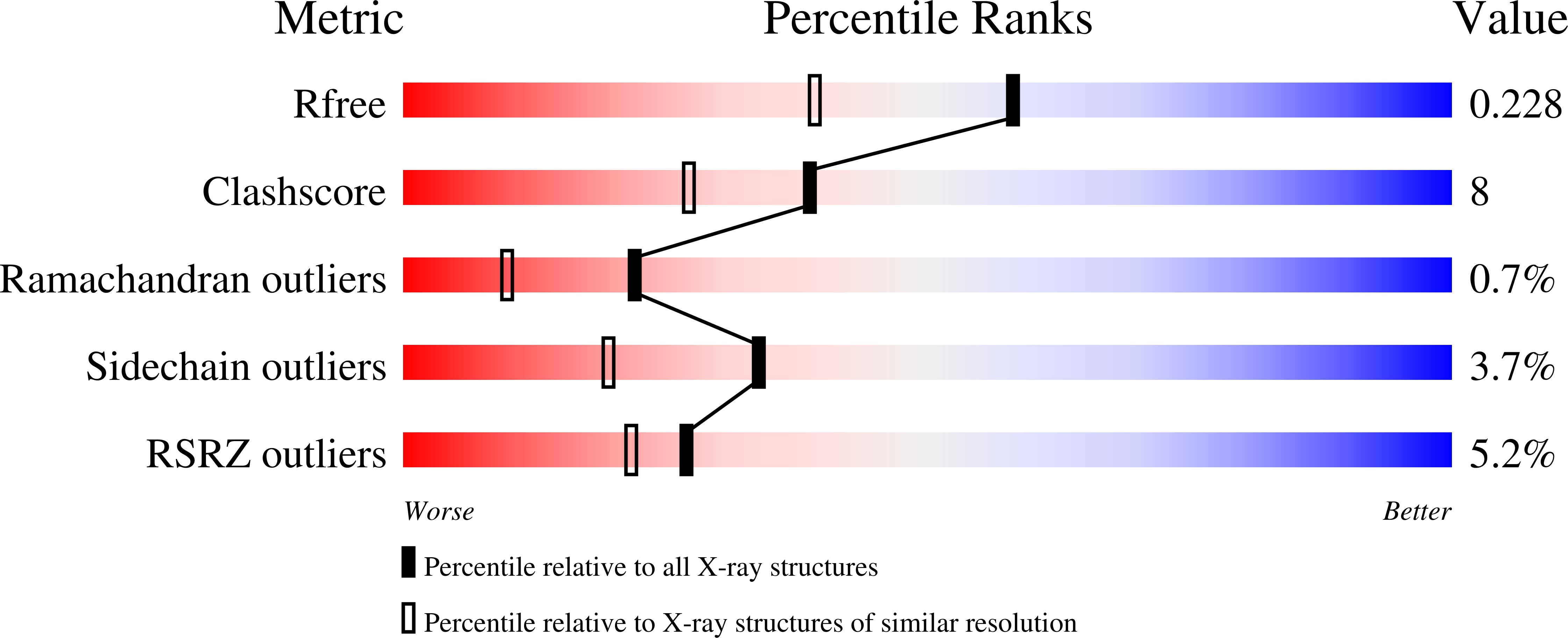
1N97

Crystal Structure of CYP175A1 from Thermus thermophillus strain HB27


Experimental Data Snapshot

  • Method: X-RAY DIFFRACTION
  • Resolution: 1.80 Å
  • R-Value Free: 0.222 
  • R-Value Work: 0.182 
  • R-Value Observed: 0.184 

wwPDB Validation   3D Report Full Report


Ligand Structure Quality Assessment 


This is version 1.4 of the entry. See complete history


Literature

Preliminary Characterization and Crystal Structure of a Thermostable Cytochrome P450 from Thermus thermophilus

Yano, J.K.Blasco, F.Li, H.Schmid, R.D.Henne, A.Poulos, T.L.

(2003) J Biol Chem 278: 608-616

  • DOI: https://doi.org/10.1074/jbc.M206568200
  • Primary Citation of Related Structures:  
    1N97

  • PubMed Abstract: 

    The second structure of a thermophile cytochrome P450, CYP175A1 from the thermophilic bacterium Thermus thermophilus HB27, has been solved to 1.8-A resolution. The overall P450 structure remains conserved despite the low sequence identity between the various P450s. The CYP175A1 structure lacks the large aromatic network found in the only other thermostable P450, CYP119, thought to contribute to thermal stability. The primary difference between CYP175A1 and its mesophile counterparts is the investment of charged residues into salt-link networks at the expense of single charge-charge interactions. Additional factors involved in the thermal stability increase are a decrease in the overall size, especially shortening of loops and connecting regions, and a decrease in the number of labile residues such as Asn, Gln, and Cys.


  • Organizational Affiliation

    Department of Molecular Biology and Biochemistry, University of California, Irvine, California 92697-3900, USA.


Macromolecules
Find similar proteins by:  (by identity cutoff)  |  3D Structure
Entity ID: 1
MoleculeChains Sequence LengthOrganismDetailsImage
CYP175A1
A, B
389Thermus thermophilus HB27Mutation(s): 0 
UniProt
Find proteins for Q746J6 (Thermus thermophilus (strain ATCC BAA-163 / DSM 7039 / HB27))
Explore Q746J6 
Go to UniProtKB:  Q746J6
Entity Groups  
Sequence Clusters30% Identity50% Identity70% Identity90% Identity95% Identity100% Identity
UniProt GroupQ746J6
Sequence Annotations
Expand
  • Reference Sequence
Small Molecules
Ligands 3 Unique
IDChains Name / Formula / InChI Key2D Diagram3D Interactions
HEM
Query on HEM

Download Ideal Coordinates CCD File 
D [auth A],
M [auth B]
PROTOPORPHYRIN IX CONTAINING FE
C34 H32 Fe N4 O4
KABFMIBPWCXCRK-RGGAHWMASA-L
SRT
Query on SRT

Download Ideal Coordinates CCD File 
C [auth A],
L [auth B]
S,R MESO-TARTARIC ACID
C4 H6 O6
FEWJPZIEWOKRBE-XIXRPRMCSA-N
EDO
Query on EDO

Download Ideal Coordinates CCD File 
E [auth A]
F [auth A]
G [auth A]
H [auth A]
I [auth A]
E [auth A],
F [auth A],
G [auth A],
H [auth A],
I [auth A],
J [auth A],
K [auth A],
N [auth B],
O [auth B],
P [auth B],
Q [auth B],
R [auth B],
S [auth B],
T [auth B],
U [auth B],
V [auth B],
W [auth B],
X [auth B]
1,2-ETHANEDIOL
C2 H6 O2
LYCAIKOWRPUZTN-UHFFFAOYSA-N
Experimental Data & Validation

Experimental Data

  • Method: X-RAY DIFFRACTION
  • Resolution: 1.80 Å
  • R-Value Free: 0.222 
  • R-Value Work: 0.182 
  • R-Value Observed: 0.184 
  • Space Group: P 1 21 1
Unit Cell:
Length ( Å )Angle ( ˚ )
a = 64.538α = 90
b = 90.699β = 99.68
c = 74.516γ = 90
Software Package:
Software NamePurpose
HKL-2000data collection
SCALEPACKdata scaling
AMoREphasing
REFMACrefinement
HKL-2000data reduction

Structure Validation

View Full Validation Report


Ligand Structure Quality Assessment 


Entry History 

Deposition Data

Revision History  (Full details and data files)

  • Version 1.0: 2003-02-25
    Type: Initial release
  • Version 1.1: 2007-10-16
    Changes: Version format compliance
  • Version 1.2: 2011-07-13
    Changes: Advisory, Source and taxonomy, Version format compliance
  • Version 1.3: 2017-02-01
    Changes: Structure summary
  • Version 1.4: 2023-08-16
    Changes: Data collection, Database references, Derived calculations, Refinement description