


Crystal Structure of Human Inosine Monophosphate Dehydrogenase type II complexed with the MPA/NAD analog C2-MAD
Risal, D., Strickler, M.D., Goldstein, B.M.To be published.
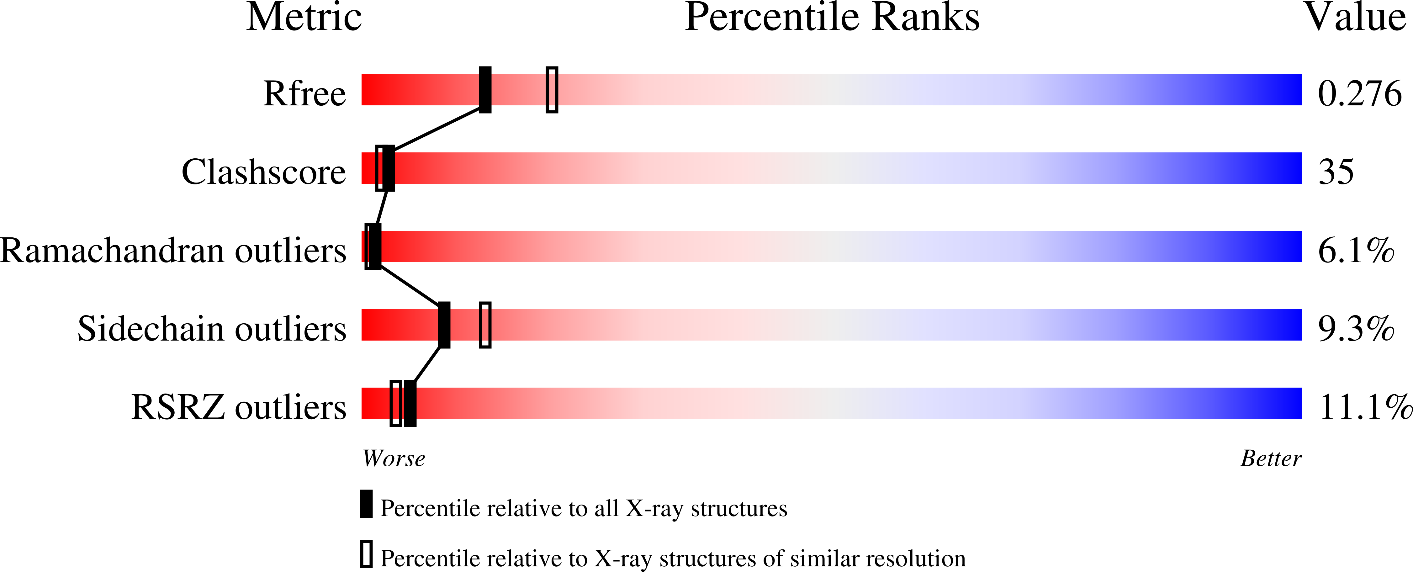
Experimental Data Snapshot
Entity ID: 1 | |||||
|---|---|---|---|---|---|
| Molecule | Chains | Sequence Length | Organism | Details | Image |
| Inosine-5'-monophosphate dehydrogenase 2 | 514 | Homo sapiens | Mutation(s): 0 Gene Names: IMPDH2 EC: 1.1.1.205 |  | |
UniProt & NIH Common Fund Data Resources | |||||
Find proteins for P12268 (Homo sapiens) Explore P12268 Go to UniProtKB: P12268 | |||||
PHAROS: P12268 GTEx: ENSG00000178035 | |||||
Entity Groups | |||||
| Sequence Clusters | 30% Identity50% Identity70% Identity90% Identity95% Identity100% Identity | ||||
| UniProt Group | P12268 | ||||
Sequence AnnotationsExpand | |||||
| |||||
| Ligands 4 Unique | |||||
|---|---|---|---|---|---|
| ID | Chains | Name / Formula / InChI Key | 2D Diagram | 3D Interactions | |
| MYD Query on MYD | E [auth A], H [auth B] | {[5-(6-AMINO-PURIN-7-YL)-3,4-DIHYDROXY-TETRAHYDRO-FURAN-2-YLMETHOXY]-HYDROXY-PHOSPHORYLMETHYL}-PHOSPHONIC ACID
MONO-[2-(4-HYDROXY-6-METHOXY-7-METHYL-3-OXO-1,3-DIHYDRO-ISOBENZOFURAN-5-YL)-ETHYL] ESTER C23 H29 N5 O13 P2 AMYUZLUBFKOUEX-JKWAKEATSA-N |  | ||
| RVP Query on RVP | D [auth A], G [auth B] | RIBAVIRIN MONOPHOSPHATE C8 H13 N4 O8 P SDWIOXKHTFOULX-AFCXAGJDSA-N |  | ||
| UNK Query on UNK | I [auth B] J [auth B] K [auth B] L [auth B] M [auth B] | UNKNOWN C4 H9 N O2 QWCKQJZIFLGMSD-VKHMYHEASA-N | |||
| K Query on K | C [auth A], F [auth B] | POTASSIUM ION K NPYPAHLBTDXSSS-UHFFFAOYSA-N |  | ||
| Length ( Å ) | Angle ( ˚ ) |
|---|---|
| a = 146.58 | α = 90 |
| b = 146.58 | β = 90 |
| c = 128.92 | γ = 90 |
| Software Name | Purpose |
|---|---|
| HKL-2000 | data collection |
| SCALA | data scaling |
| MOLREP | phasing |
| CNS | refinement |
| HKL-2000 | data reduction |
| CCP4 | data scaling |
RCSB PDB (citation) is hosted by
RCSB PDB is a member of the

