1PS1

PENTALENENE SYNTHASE


Experimental Data Snapshot

  • Method: X-RAY DIFFRACTION
  • Resolution: 2.60 Å
  • R-Value Free: 0.273 
  • R-Value Work: 0.198 
  • R-Value Observed: 0.198 

wwPDB Validation   3D Report Full Report


This is version 1.2 of the entry. See complete history


Literature

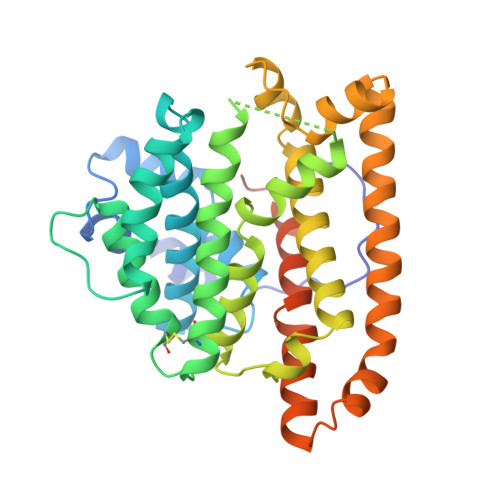
Crystal structure of pentalenene synthase: mechanistic insights on terpenoid cyclization reactions in biology.

Lesburg, C.A.Zhai, G.Cane, D.E.Christianson, D.W.

(1997) Science 277: 1820-1824

  • DOI: https://doi.org/10.1126/science.277.5333.1820
  • Primary Citation of Related Structures:  
    1PS1

  • PubMed Abstract: 

    The crystal structure of pentalenene synthase at 2.6 angstrom resolution reveals critical active site features responsible for the cyclization of farnesyl diphosphate into the tricyclic hydrocarbon pentalenene. Metal-triggered substrate ionization initiates catalysis, and the alpha-barrel active site serves as a template to channel and stabilize the conformations of reactive carbocation intermediates through a complex cyclization cascade. The core active site structure of the enzyme may be preserved among the greater family of terpenoid synthases, possibly implying divergence from a common ancestral synthase to satisfy biological requirements for increasingly diverse natural products.


  • Organizational Affiliation

    Roy and Diana Vagelos Laboratories, Department of Chemistry, University of Pennsylvania, Philadelphia, PA 19104-6323, USA.


Macromolecules
Find similar proteins by:  (by identity cutoff)  |  3D Structure
Entity ID: 1
MoleculeChains Sequence LengthOrganismDetailsImage
PENTALENENE SYNTHASE
A, B
337Streptomyces exfoliatusMutation(s): 0 
EC: 4.6.1.5
UniProt
Find proteins for Q55012 (Streptomyces exfoliatus)
Explore Q55012 
Go to UniProtKB:  Q55012
Entity Groups  
Sequence Clusters30% Identity50% Identity70% Identity90% Identity95% Identity100% Identity
UniProt GroupQ55012
Sequence Annotations
Expand
  • Reference Sequence
Small Molecules
Ligands 1 Unique
IDChains Name / Formula / InChI Key2D Diagram3D Interactions
PBM
Query on PBM

Download Ideal Coordinates CCD File 
C [auth A]TRIMETHYL LEAD ION
C3 H9 Pb
SNBFOEGVGALPSE-UHFFFAOYSA-N
Experimental Data & Validation

Experimental Data

  • Method: X-RAY DIFFRACTION
  • Resolution: 2.60 Å
  • R-Value Free: 0.273 
  • R-Value Work: 0.198 
  • R-Value Observed: 0.198 
  • Space Group: P 63
Unit Cell:
Length ( Å )Angle ( ˚ )
a = 178.72α = 90
b = 178.72β = 90
c = 56.62γ = 120
Software Package:
Software NamePurpose
DENZOdata reduction
SCALEPACKdata scaling
SOLOMONphasing
X-PLORrefinement

Structure Validation

View Full Validation Report


Entry History 

Deposition Data

Revision History  (Full details and data files)

  • Version 1.0: 1998-03-25
    Type: Initial release
  • Version 1.1: 2008-03-24
    Changes: Version format compliance
  • Version 1.2: 2011-07-13
    Changes: Source and taxonomy, Version format compliance