1QGI

CHITOSANASE FROM BACILLUS CIRCULANS


Experimental Data Snapshot

  • Method: X-RAY DIFFRACTION
  • Resolution: 1.60 Å
  • R-Value Free: 0.235 
  • R-Value Work: 0.192 

wwPDB Validation   3D Report Full Report


This is version 2.1 of the entry. See complete history


Literature

Crystal structure of chitosanase from Bacillus circulans MH-K1 at 1.6-A resolution and its substrate recognition mechanism.

Saito, J.Kita, A.Higuchi, Y.Nagata, Y.Ando, A.Miki, K.

(1999) J Biol Chem 274: 30818-30825

  • DOI: https://doi.org/10.1074/jbc.274.43.30818
  • Primary Citation of Related Structures:  
    1QGI

  • PubMed Abstract: 

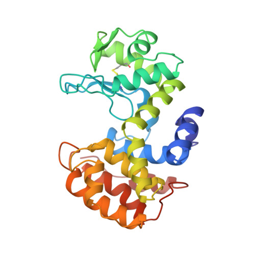
    Chitosanase from Bacillus circulans MH-K1 is a 29-kDa extracellular protein composed of 259 amino acids. The crystal structure of chitosanase from B. circulans MH-K1 has been determined by multiwavelength anomalous diffraction method and refined to crystallographic R = 19.2% (R(free) = 23.5%) for the diffraction data at 1.6-A resolution collected by synchrotron radiation. The enzyme has two globular upper and lower domains, which generate the active site cleft for the substrate binding. The overall molecular folding is similar to chitosanase from Streptomyces sp. N174, although there is only 20% identity at the amino acid sequence level between both chitosanases. However, there are three regions in which the topology is remarkably different. In addition, the disulfide bridge between Cys(50) and Cys(124) joins the beta1 strand and the alpha7 helix, which is not conserved among other chitosanases. The orientation of two backbone helices, which connect the two domains, is also different and is responsible for the differences in size and shape of the active site cleft in these two chitosanases. This structural difference in the active site cleft is the reason why the enzymes specifically recognize different substrates and catalyze different types of chitosan degradation.


  • Organizational Affiliation

    Department of Chemistry, Graduate School of Science, Kyoto University, Sakyo-ku, Kyoto 606-8502, Japan.


Macromolecules
Find similar proteins by:  (by identity cutoff)  |  3D Structure
Entity ID: 1
MoleculeChains Sequence LengthOrganismDetailsImage
PROTEIN (CHITOSANASE)259Niallia circulansMutation(s): 0 
EC: 3.2.1.132
UniProt
Find proteins for P33673 (Niallia circulans)
Explore P33673 
Go to UniProtKB:  P33673
Entity Groups  
Sequence Clusters30% Identity50% Identity70% Identity90% Identity95% Identity100% Identity
UniProt GroupP33673
Sequence Annotations
Expand
  • Reference Sequence
Oligosaccharides

Help

Entity ID: 2
MoleculeChains Length2D Diagram Glycosylation3D Interactions
2-amino-2-deoxy-beta-D-glucopyranose-(1-4)-2-amino-2-deoxy-beta-D-glucopyranose-(1-4)-2-acetamido-2-deoxy-beta-D-glucopyranose
B
3N/A
Glycosylation Resources
GlyTouCan:  G93015JH
GlyCosmos:  G93015JH
Small Molecules
Ligands 1 Unique
IDChains Name / Formula / InChI Key2D Diagram3D Interactions
SO4
Query on SO4

Download Ideal Coordinates CCD File 
C [auth A]SULFATE ION
O4 S
QAOWNCQODCNURD-UHFFFAOYSA-L
Experimental Data & Validation

Experimental Data

  • Method: X-RAY DIFFRACTION
  • Resolution: 1.60 Å
  • R-Value Free: 0.235 
  • R-Value Work: 0.192 
  • Space Group: P 21 21 2
Unit Cell:
Length ( Å )Angle ( ˚ )
a = 43.3α = 90
b = 128β = 90
c = 57.7γ = 90
Software Package:
Software NamePurpose
X-PLORrefinement

Structure Validation

View Full Validation Report


Entry History 

Deposition Data

Revision History  (Full details and data files)

  • Version 1.0: 1999-10-25
    Type: Initial release
  • Version 1.1: 2008-04-26
    Changes: Version format compliance
  • Version 1.2: 2011-07-13
    Changes: Non-polymer description, Version format compliance
  • Version 2.0: 2020-07-29
    Type: Remediation
    Reason: Carbohydrate remediation
    Changes: Advisory, Atomic model, Data collection, Database references, Derived calculations, Structure summary
  • Version 2.1: 2023-12-27
    Changes: Data collection, Database references, Structure summary