1XXN

Crystal structure of a mesophilic xylanase A from Bacillus subtilis 1A1


Experimental Data Snapshot

  • Method: X-RAY DIFFRACTION
  • Resolution: 1.70 Å
  • R-Value Free: 0.212 
  • R-Value Work: 0.171 
  • R-Value Observed: 0.173 

wwPDB Validation   3D Report Full Report


Ligand Structure Quality Assessment 


This is version 1.5 of the entry. See complete history


Literature

Correlation of temperature induced conformation change with optimum catalytic activity in the recombinant G/11 xylanase A from Bacillus subtilis strain 168 (1A1).

Murakami, M.T.Arni, R.K.Vieira, D.S.Degreve, L.Ruller, R.Ward, R.J.

(2005) FEBS Lett 579: 6505-6510

  • DOI: https://doi.org/10.1016/j.febslet.2005.10.039
  • Primary Citation of Related Structures:  
    1XXN

  • PubMed Abstract: 

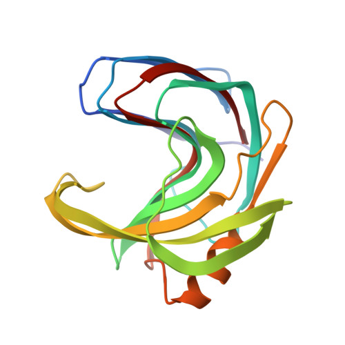
    The 1.7A resolution crystal structure of recombinant family G/11 beta-1,4-xylanase (rXynA) from Bacillus subtilis 1A1 shows a jellyroll fold in which two curved beta-sheets form the active-site and substrate-binding cleft. The onset of thermal denaturation of rXynA occurs at 328 K, in excellent agreement with the optimum catalytic temperature. Molecular dynamics simulations at temperatures of 298-328 K demonstrate that below the optimum temperature the thumb loop and palm domain adopt a closed conformation. However, at 328 K these two domains separate facilitating substrate access to the active-site pocket, thereby accounting for the optimum catalytic temperature of the rXynA.


  • Organizational Affiliation

    Department of Physics, IBILCE/UNESP, Cristovão Colombo 2265, São José do Rio Preto, São Paulo, Brazil.


Macromolecules
Find similar proteins by:  (by identity cutoff)  |  3D Structure
Entity ID: 1
MoleculeChains Sequence LengthOrganismDetailsImage
Endo-1,4-beta-xylanase A185Bacillus subtilisMutation(s): 0 
Gene Names: xynA
EC: 3.2.1.8
UniProt
Find proteins for P18429 (Bacillus subtilis (strain 168))
Explore P18429 
Go to UniProtKB:  P18429
Entity Groups  
Sequence Clusters30% Identity50% Identity70% Identity90% Identity95% Identity100% Identity
UniProt GroupP18429
Sequence Annotations
Expand
  • Reference Sequence
Small Molecules
Ligands 1 Unique
IDChains Name / Formula / InChI Key2D Diagram3D Interactions
SRT
Query on SRT

Download Ideal Coordinates CCD File 
B [auth A]S,R MESO-TARTARIC ACID
C4 H6 O6
FEWJPZIEWOKRBE-XIXRPRMCSA-N
Experimental Data & Validation

Experimental Data

  • Method: X-RAY DIFFRACTION
  • Resolution: 1.70 Å
  • R-Value Free: 0.212 
  • R-Value Work: 0.171 
  • R-Value Observed: 0.173 
  • Space Group: P 21 21 2
Unit Cell:
Length ( Å )Angle ( ˚ )
a = 50.931α = 90
b = 70.499β = 90
c = 42.044γ = 90
Software Package:
Software NamePurpose
REFMACrefinement
MAR345data collection
SCALEPACKdata scaling
AMoREphasing

Structure Validation

View Full Validation Report


Ligand Structure Quality Assessment 


Entry History 

Deposition Data

Revision History  (Full details and data files)

  • Version 1.0: 2005-10-18
    Type: Initial release
  • Version 1.1: 2008-04-30
    Changes: Version format compliance
  • Version 1.2: 2011-07-13
    Changes: Version format compliance
  • Version 1.3: 2013-03-06
    Changes: Other
  • Version 1.4: 2017-10-11
    Changes: Refinement description
  • Version 1.5: 2023-08-23
    Changes: Data collection, Database references, Derived calculations, Refinement description