2WHP

Crystal structure of acetylcholinesterase, phosphonylated by sarin and in complex with HI-6


Experimental Data Snapshot

  • Method: X-RAY DIFFRACTION
  • Resolution: 2.20 Å
  • R-Value Free: 0.210 
  • R-Value Work: 0.175 
  • R-Value Observed: 0.176 

wwPDB Validation   3D Report Full Report


Ligand Structure Quality Assessment 


This is version 1.5 of the entry. See complete history


Literature

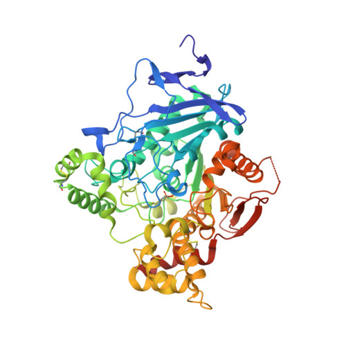
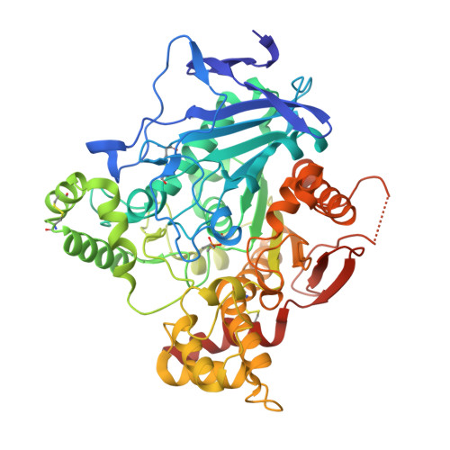
Structure of Hi-6Sarin-Acetylcholinesterase Determined by X-Ray Crystallography and Molecular Dynamics Simulation: Reactivator Mechanism and Design.

Ekstrom, F.Hornberg, A.Artursson, E.Hammarstrom, L.G.Schneider, G.Pang, Y.P.

(2009) PLoS One 4: E5957

  • DOI: https://doi.org/10.1371/journal.pone.0005957
  • Primary Citation of Related Structures:  
    2WHP, 2WHR

  • PubMed Abstract: 

    Organophosphonates such as isopropyl metylphosphonofluoridate (sarin) are extremely toxic as they phosphonylate the catalytic serine residue of acetylcholinesterase (AChE), an enzyme essential to humans and other species. Design of effective AChE reactivators as antidotes to various organophosphonates requires information on how the reactivators interact with the phosphonylated AChEs. However, such information has not been available hitherto because of three main challenges. First, reactivators are generally flexible in order to change from the ground state to the transition state for reactivation; this flexibility discourages determination of crystal structures of AChE in complex with effective reactivators that are intrinsically disordered. Second, reactivation occurs upon binding of a reactivator to the phosphonylated AChE. Third, the phosphorous conjugate can develop resistance to reactivation. We have identified crystallographic conditions that led to the determination of a crystal structure of the sarin(nonaged)-conjugated mouse AChE in complex with [(E)-[1-[(4-carbamoylpyridin-1-ium-1-yl)methoxymethyl]pyridin-2-ylidene]methyl]-oxoazanium dichloride (HI-6) at a resolution of 2.2 A. In this structure, the carboxyamino-pyridinium ring of HI-6 is sandwiched by Tyr124 and Trp286, however, the oxime-pyridinium ring is disordered. By combining crystallography with microsecond molecular dynamics simulation, we determined the oxime-pyridinium ring structure, which shows that the oxime group of HI-6 can form a hydrogen-bond network to the sarin isopropyl ether oxygen, and a water molecule is able to form a hydrogen bond to the catalytic histidine residue and subsequently deprotonates the oxime for reactivation. These results offer insights into the reactivation mechanism of HI-6 and design of better reactivators.


  • Organizational Affiliation

    Swedish Defence Research Agency, CBRN Defence and Security, Umeå, Sweden. fredrik.ekstrom@foi.se


Macromolecules
Find similar proteins by:  (by identity cutoff)  |  3D Structure
Entity ID: 1
MoleculeChains Sequence LengthOrganismDetailsImage
ACETYLCHOLINESTERASE548Mus musculusMutation(s): 0 
EC: 3.1.1.7
UniProt & NIH Common Fund Data Resources
Find proteins for P21836 (Mus musculus)
Explore P21836 
Go to UniProtKB:  P21836
IMPC:  MGI:87876
Entity Groups  
Sequence Clusters30% Identity50% Identity70% Identity90% Identity95% Identity100% Identity
UniProt GroupP21836
Sequence Annotations
Expand
  • Reference Sequence
Find similar proteins by:  (by identity cutoff)  |  3D Structure
Entity ID: 2
MoleculeChains Sequence LengthOrganismDetailsImage
ACETYLCHOLINESTERASE548Mus musculusMutation(s): 0 
EC: 3.1.1.7
UniProt & NIH Common Fund Data Resources
Find proteins for P21836 (Mus musculus)
Explore P21836 
Go to UniProtKB:  P21836
IMPC:  MGI:87876
Entity Groups  
Sequence Clusters30% Identity50% Identity70% Identity90% Identity95% Identity100% Identity
UniProt GroupP21836
Sequence Annotations
Expand
  • Reference Sequence
Small Molecules
Ligands 5 Unique
IDChains Name / Formula / InChI Key2D Diagram3D Interactions
HI6
Query on HI6

Download Ideal Coordinates CCD File 
E [auth A],
H [auth B]
4-(AMINOCARBONYL)-1-[({2-[(E)-(HYDROXYIMINO)METHYL]PYRIDINIUM-1-YL}METHOXY)METHYL]PYRIDINIUM
C14 H16 N4 O3
FJZDLOMCEPUCII-UHFFFAOYSA-P
P6G
Query on P6G

Download Ideal Coordinates CCD File 
J [auth B]HEXAETHYLENE GLYCOL
C12 H26 O7
IIRDTKBZINWQAW-UHFFFAOYSA-N
NAG
Query on NAG

Download Ideal Coordinates CCD File 
C [auth A],
D [auth A],
G [auth B]
2-acetamido-2-deoxy-beta-D-glucopyranose
C8 H15 N O6
OVRNDRQMDRJTHS-FMDGEEDCSA-N
PEG
Query on PEG

Download Ideal Coordinates CCD File 
I [auth B]DI(HYDROXYETHYL)ETHER
C4 H10 O3
MTHSVFCYNBDYFN-UHFFFAOYSA-N
CO3
Query on CO3

Download Ideal Coordinates CCD File 
F [auth A]CARBONATE ION
C O3
BVKZGUZCCUSVTD-UHFFFAOYSA-L
Modified Residues  1 Unique
IDChains TypeFormula2D DiagramParent
SGB
Query on SGB
A
L-PEPTIDE LINKINGC7 H16 N O5 PSER
Binding Affinity Annotations 
IDSourceBinding Affinity
HI6 BindingDB:  2WHP Kd: min: 1.50e+4, max: 2.34e+5 (nM) from 6 assay(s)
IC50: min: 6.36e+5, max: 1.36e+6 (nM) from 3 assay(s)
PDBBind:  2WHP Kd: 1.00e+5 (nM) from 1 assay(s)
Binding MOAD:  2WHP Kd: 1.00e+5 (nM) from 1 assay(s)
Experimental Data & Validation

Experimental Data

  • Method: X-RAY DIFFRACTION
  • Resolution: 2.20 Å
  • R-Value Free: 0.210 
  • R-Value Work: 0.175 
  • R-Value Observed: 0.176 
  • Space Group: P 21 21 21
Unit Cell:
Length ( Å )Angle ( ˚ )
a = 79.26α = 90
b = 112.15β = 90
c = 226.95γ = 90
Software Package:
Software NamePurpose
PHENIXrefinement
XDSdata reduction
SCALAdata scaling
REFMACphasing

Structure Validation

View Full Validation Report


Ligand Structure Quality Assessment 


Entry History 

Deposition Data

Revision History  (Full details and data files)

  • Version 1.0: 2009-06-30
    Type: Initial release
  • Version 1.1: 2011-05-08
    Changes: Version format compliance
  • Version 1.2: 2011-07-13
    Changes: Version format compliance
  • Version 1.3: 2018-01-17
    Changes: Data collection
  • Version 1.4: 2020-07-29
    Type: Remediation
    Reason: Carbohydrate remediation
    Changes: Data collection, Derived calculations, Other, Structure summary
  • Version 1.5: 2023-12-13
    Changes: Data collection, Database references, Refinement description, Structure summary