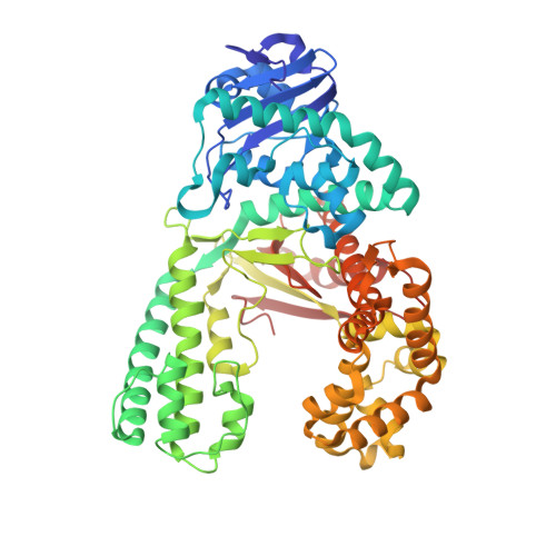
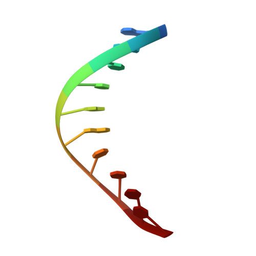
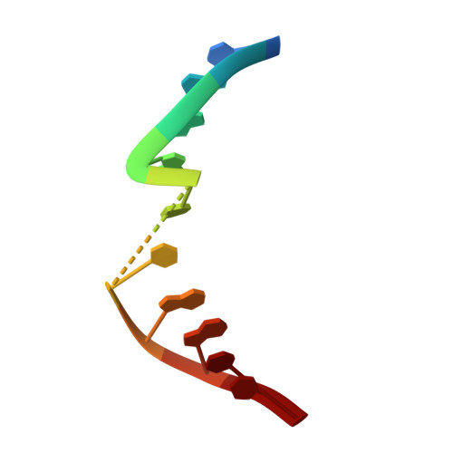
2Y1I

Crystal structure of a S-diastereomer analogue of the spore photoproduct in complex with fragment DNA polymerase I from Bacillus stearothermophilus


Experimental Data Snapshot

  • Method: X-RAY DIFFRACTION
  • Resolution: 2.78 Å
  • R-Value Free: 0.273 
  • R-Value Work: 0.226 
  • R-Value Observed: 0.228 

wwPDB Validation   3D Report Full Report


This is version 1.2 of the entry. See complete history


Literature

Crystal Structures and Repair Studies Reveal the Identity and the Base-Pairing Properties of the Uv-Induced Spore Photoproduct DNA Lesion.

Heil, K.Kneuttinger, A.C.Schneider, S.Lischke, U.Carell, T.

(2011) Chemistry 17: 9651

  • DOI: https://doi.org/10.1002/chem.201100177
  • Primary Citation of Related Structures:  
    2Y1I, 2Y1J

  • PubMed Abstract: 

    UV light is one of the major causes of DNA damage. In spore DNA, due to an unusual packing of the genetic material, a special spore photoproduct lesion (SP lesion) is formed, which is repaired by the enzyme spore photoproduct lyase (Spl), a radical S-adenosylmethionine (SAM) enzyme. We report here the synthesis and DNA incorporation of a DNA SP lesion analogue lacking the phosphodiester backbone. The oligonucleotides were used for repair studies and they were cocrystallized with a polymerase enzyme as a template to clarify the configuration of the SP lesion and to provide information about the base-pairing properties of the lesion. The structural analysis together with repair studies allowed us to clarify the identity of the preferentially repaired lesion diastereoisomer.


  • Organizational Affiliation

    Center for Integrated Protein Science (CiPSM) at the Department of Chemistry, Ludwig-Maximilians-University, Munich, Germany.


Macromolecules

Find similar proteins by:  (by identity cutoff)  |  3D Structure
Entity ID: 1
MoleculeChains Sequence LengthOrganismDetailsImage
DNA POLYMERASE I580Geobacillus stearothermophilusMutation(s): 0 
EC: 2.7.7.7
UniProt
Find proteins for E1C9K5 (Geobacillus stearothermophilus)
Explore E1C9K5 
Go to UniProtKB:  E1C9K5
Entity Groups  
Sequence Clusters30% Identity50% Identity70% Identity90% Identity95% Identity100% Identity
UniProt GroupE1C9K5
Sequence Annotations
Expand
  • Reference Sequence

Find similar nucleic acids by:  Sequence   |   3D Structure  

Entity ID: 2
MoleculeChains LengthOrganismImage
5'-D(*GP*AP*CP*CP*AP*AP*CP*CP*CP*TP)-3'10synthetic construct
Sequence Annotations
Expand
  • Reference Sequence

Find similar nucleic acids by:  Sequence   |   3D Structure  

Entity ID: 3
MoleculeChains LengthOrganismImage
5'-D(*AP*GP*GP*GP*PBTP*THM*GP*GP*TP*CP)-3'10synthetic construct
Sequence Annotations
Expand
  • Reference Sequence
Experimental Data & Validation

Experimental Data

  • Method: X-RAY DIFFRACTION
  • Resolution: 2.78 Å
  • R-Value Free: 0.273 
  • R-Value Work: 0.226 
  • R-Value Observed: 0.228 
  • Space Group: P 21 21 21
Unit Cell:
Length ( Å )Angle ( ˚ )
a = 88.669α = 90
b = 93.793β = 90
c = 107.101γ = 90
Software Package:
Software NamePurpose
REFMACrefinement
XDSdata reduction
SCALAdata scaling
PHASERphasing

Structure Validation

View Full Validation Report


Entry History 

Deposition Data

Revision History  (Full details and data files)

  • Version 1.0: 2011-07-27
    Type: Initial release
  • Version 1.1: 2011-11-02
    Changes: Database references
  • Version 1.2: 2023-12-20
    Changes: Data collection, Database references, Derived calculations, Other, Refinement description