3M8Z

Phosphopentomutase from Bacillus cereus bound with ribose-5-phosphate


Experimental Data Snapshot

  • Method: X-RAY DIFFRACTION
  • Resolution: 1.80 Å
  • R-Value Free: 0.198 
  • R-Value Work: 0.168 
  • R-Value Observed: 0.170 

wwPDB Validation   3D Report Full Report


Ligand Structure Quality Assessment 


This is version 2.1 of the entry. See complete history


Literature

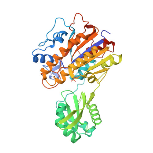
Bacillus cereus Phosphopentomutase Is an Alkaline Phosphatase Family Member That Exhibits an Altered Entry Point into the Catalytic Cycle.

Panosian, T.D.Nannemann, D.P.Watkins, G.R.Phelan, V.V.McDonald, W.H.Wadzinski, B.E.Bachmann, B.O.Iverson, T.M.

(2011) J Biol Chem 286: 8043-8054

  • DOI: https://doi.org/10.1074/jbc.M110.201350
  • Primary Citation of Related Structures:  
    3M8W, 3M8Y, 3M8Z, 3OT9

  • PubMed Abstract: 

    Bacterial phosphopentomutases (PPMs) are alkaline phosphatase superfamily members that interconvert α-D-ribose 5-phosphate (ribose 5-phosphate) and α-D-ribose 1-phosphate (ribose 1-phosphate). We investigated the reaction mechanism of Bacillus cereus PPM using a combination of structural and biochemical studies. Four high resolution crystal structures of B. cereus PPM revealed the active site architecture, identified binding sites for the substrate ribose 5-phosphate and the activator α-D-glucose 1,6-bisphosphate (glucose 1,6-bisphosphate), and demonstrated that glucose 1,6-bisphosphate increased phosphorylation of the active site residue Thr-85. The phosphorylation of Thr-85 was confirmed by Western and mass spectroscopic analyses. Biochemical assays identified Mn(2+)-dependent enzyme turnover and demonstrated that glucose 1,6-bisphosphate treatment increases enzyme activity. These results suggest that protein phosphorylation activates the enzyme, which supports an intermolecular transferase mechanism. We confirmed intermolecular phosphoryl transfer using an isotope relay assay in which PPM reactions containing mixtures of ribose 5-[(18)O(3)]phosphate and [U-(13)C(5)]ribose 5-phosphate were analyzed by mass spectrometry. This intermolecular phosphoryl transfer is seemingly counter to what is anticipated from phosphomutases employing a general alkaline phosphatase reaction mechanism, which are reported to catalyze intramolecular phosphoryl transfer. However, the two mechanisms may be reconciled if substrate encounters the enzyme at a different point in the catalytic cycle.


  • Organizational Affiliation

    From the Departments of Pharmacology and.


Macromolecules
Find similar proteins by:  (by identity cutoff)  |  3D Structure
Entity ID: 1
MoleculeChains Sequence LengthOrganismDetailsImage
Phosphopentomutase
A, B, C
399Bacillus cereus ATCC 14579Mutation(s): 0 
Gene Names: deoBBC_4087
EC: 5.4.2.7
UniProt
Find proteins for Q818Z9 (Bacillus cereus (strain ATCC 14579 / DSM 31 / CCUG 7414 / JCM 2152 / NBRC 15305 / NCIMB 9373 / NCTC 2599 / NRRL B-3711))
Explore Q818Z9 
Go to UniProtKB:  Q818Z9
Entity Groups  
Sequence Clusters30% Identity50% Identity70% Identity90% Identity95% Identity100% Identity
UniProt GroupQ818Z9
Sequence Annotations
Expand
  • Reference Sequence
Small Molecules
Ligands 5 Unique
IDChains Name / Formula / InChI Key2D Diagram3D Interactions
HSX
Query on HSX

Download Ideal Coordinates CCD File 
J [auth A],
K [auth A]
5-O-phosphono-alpha-D-ribofuranose
C5 H11 O8 P
KTVPXOYAKDPRHY-AIHAYLRMSA-N
TRS
Query on TRS

Download Ideal Coordinates CCD File 
G [auth A],
O [auth B]
2-AMINO-2-HYDROXYMETHYL-PROPANE-1,3-DIOL
C4 H12 N O3
LENZDBCJOHFCAS-UHFFFAOYSA-O
GOL
Query on GOL

Download Ideal Coordinates CCD File 
I [auth A],
P [auth B]
GLYCEROL
C3 H8 O3
PEDCQBHIVMGVHV-UHFFFAOYSA-N
ACT
Query on ACT

Download Ideal Coordinates CCD File 
H [auth A]ACETATE ION
C2 H3 O2
QTBSBXVTEAMEQO-UHFFFAOYSA-M
MN
Query on MN

Download Ideal Coordinates CCD File 
D [auth A]
E [auth A]
F [auth A]
L [auth B]
M [auth B]
D [auth A],
E [auth A],
F [auth A],
L [auth B],
M [auth B],
N [auth B],
Q [auth C],
R [auth C],
S [auth C]
MANGANESE (II) ION
Mn
WAEMQWOKJMHJLA-UHFFFAOYSA-N
Modified Residues  1 Unique
IDChains TypeFormula2D DiagramParent
TPO
Query on TPO
A, B, C
L-PEPTIDE LINKINGC4 H10 N O6 PTHR
Experimental Data & Validation

Experimental Data

  • Method: X-RAY DIFFRACTION
  • Resolution: 1.80 Å
  • R-Value Free: 0.198 
  • R-Value Work: 0.168 
  • R-Value Observed: 0.170 
  • Space Group: P 1 21 1
Unit Cell:
Length ( Å )Angle ( ˚ )
a = 91.588α = 90
b = 76.755β = 108.66
c = 107.083γ = 90
Software Package:
Software NamePurpose
REFMACrefinement
CNSrefinement
HKL-2000data reduction
SCALEPACKdata scaling
CNSphasing

Structure Validation

View Full Validation Report


Ligand Structure Quality Assessment 


Entry History 

Deposition Data

Revision History  (Full details and data files)

  • Version 1.0: 2010-12-29
    Type: Initial release
  • Version 1.1: 2011-07-13
    Changes: Version format compliance
  • Version 1.2: 2017-05-31
    Changes: Database references
  • Version 1.3: 2019-07-17
    Changes: Data collection, Derived calculations, Refinement description
  • Version 2.0: 2020-07-29
    Type: Remediation
    Reason: Carbohydrate remediation
    Changes: Atomic model, Data collection, Derived calculations
  • Version 2.1: 2023-09-06
    Changes: Advisory, Data collection, Database references, Refinement description, Structure summary