3QDK

Structural insight on mechanism and diverse substrate selection strategy of ribulokinase


Experimental Data Snapshot

  • Method: X-RAY DIFFRACTION
  • Resolution: 2.31 Å
  • R-Value Free: 0.274 
  • R-Value Work: 0.224 
  • R-Value Observed: 0.224 

wwPDB Validation   3D Report Full Report


Ligand Structure Quality Assessment 


This is version 1.5 of the entry. See complete history


Literature

Structural insight into mechanism and diverse substrate selection strategy of L-ribulokinase.

Agarwal, R.Burley, S.K.Swaminathan, S.

(2012) Proteins 80: 261-268

  • DOI: https://doi.org/10.1002/prot.23202
  • Primary Citation of Related Structures:  
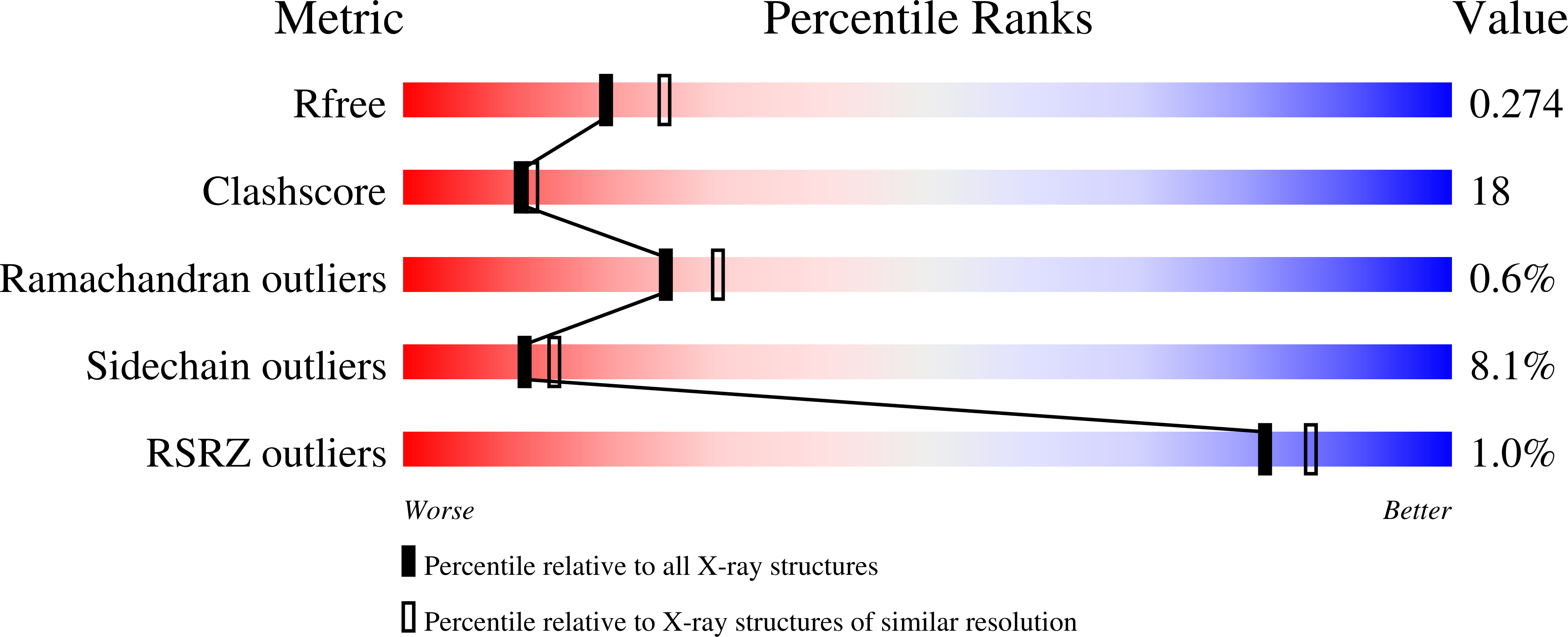
    3QDK

  • PubMed Abstract: 

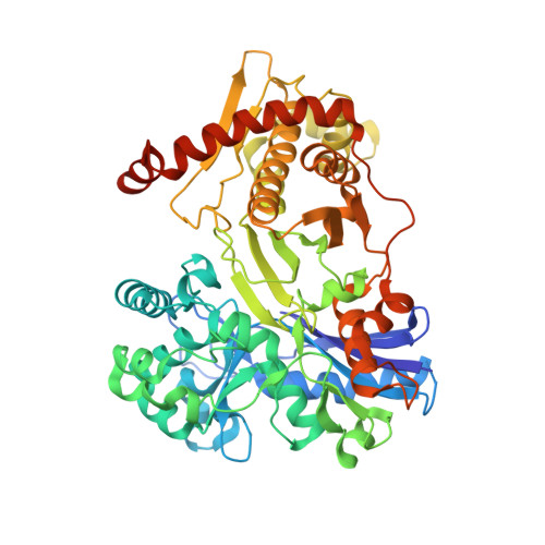
    The araBAD operon encodes three different enzymes required for catabolism of L-arabinose, which is one of the most abundant monosaccharides in nature. L-ribulokinase, encoded by the araB gene, catalyzes conversion of L-ribulose to L-ribulose-5-phosphate, the second step in the catabolic pathway. Unlike other kinases, ribulokinase exhibits diversity in substrate selectivity and catalyzes phosphorylation of all four 2-ketopentose sugars with comparable k(cat) values. To understand ribulokinase recognition and phosphorylation of a diverse set of substrates, we have determined the X-ray structure of ribulokinase from Bacillus halodurans bound to L-ribulose and investigated its substrate and ATP co-factor binding properties. The polypeptide chain is folded into two domains, one small and the other large, with a deep cleft in between. By analogy with related sugar kinases, we identified (447)GGLPQK(452) as the ATP-binding motif within the smaller domain. L-ribulose binds in the cleft between the two domains via hydrogen bonds with the side chains of highly conserved Trp126, Lys208, Asp274, and Glu329 and the main chain nitrogen of Ala96. The interaction of L-ribulokinase with L-ribulose reveals versatile structural features that help explain recognition of various 2-ketopentose substrates and competitive inhibition by L-erythrulose. Comparison of our structure to that of the structures of other sugar kinases revealed conformational variations that suggest domain-domain closure movements are responsible for establishing the observed active site environment.


  • Organizational Affiliation

    Biology Department, Brookhaven National Laboratory, Upton, New York 11973, USA.


Macromolecules
Find similar proteins by:  (by identity cutoff)  |  3D Structure
Entity ID: 1
MoleculeChains Sequence LengthOrganismDetailsImage
Ribulokinase
A, B, C, D
572Halalkalibacterium haloduransMutation(s): 0 
Gene Names: araBBH1872
EC: 2.7.1.16
UniProt
Find proteins for Q9KBQ3 (Halalkalibacterium halodurans (strain ATCC BAA-125 / DSM 18197 / FERM 7344 / JCM 9153 / C-125))
Explore Q9KBQ3 
Go to UniProtKB:  Q9KBQ3
Entity Groups  
Sequence Clusters30% Identity50% Identity70% Identity90% Identity95% Identity100% Identity
UniProt GroupQ9KBQ3
Sequence Annotations
Expand
  • Reference Sequence
Small Molecules
Ligands 1 Unique
IDChains Name / Formula / InChI Key2D Diagram3D Interactions
QDK
Query on QDK

Download Ideal Coordinates CCD File 
E [auth A]L-ribulose
C5 H10 O5
ZAQJHHRNXZUBTE-UCORVYFPSA-N
Experimental Data & Validation

Experimental Data

  • Method: X-RAY DIFFRACTION
  • Resolution: 2.31 Å
  • R-Value Free: 0.274 
  • R-Value Work: 0.224 
  • R-Value Observed: 0.224 
  • Space Group: P 1 21 1
Unit Cell:
Length ( Å )Angle ( ˚ )
a = 56.683α = 90
b = 88.84β = 92.75
c = 230.525γ = 90
Software Package:
Software NamePurpose
CBASSdata collection
SHELXDphasing
SHARPphasing
CNSrefinement
Cootmodel building
CCP4refinement
HKL-2000data reduction
SCALEPACKdata scaling

Structure Validation

View Full Validation Report


Ligand Structure Quality Assessment 


Entry History 

Deposition Data

Revision History  (Full details and data files)

  • Version 1.0: 2011-02-09
    Type: Initial release
  • Version 1.1: 2011-07-13
    Changes: Version format compliance
  • Version 1.2: 2011-09-28
    Changes: Database references
  • Version 1.3: 2011-12-28
    Changes: Database references
  • Version 1.4: 2021-02-10
    Changes: Database references, Derived calculations, Structure summary
  • Version 1.5: 2024-02-21
    Changes: Data collection, Database references