3ZGG

Crystal structure of the Fucosylgalactoside alpha N- acetylgalactosaminyltransferase (GTA, cisAB mutant L266G, G268A) in complex with NPE caged UDP-Gal (C222(1) space group)


Experimental Data Snapshot

  • Method: X-RAY DIFFRACTION
  • Resolution: 1.90 Å
  • R-Value Free: 0.226 
  • R-Value Work: 0.184 
  • R-Value Observed: 0.186 

wwPDB Validation   3D Report Full Report


Ligand Structure Quality Assessment 


This is version 1.3 of the entry. See complete history


Literature

Structures of a Human Blood Group Glycosyltransferase in Complex with a Photo-Activatable Udp-Gal Derivative Reveal Two Different Binding Conformations

Jorgensen, R.Batot, G.Mannerstedt, K.Imberty, A.Breton, C.Hindsgaul, O.Royant, A.Palcic, M.M.

(2014) Acta Crystallogr Sect F Struct Biol Cryst Commun 70: 1015

  • DOI: https://doi.org/10.1107/S2053230X1401259X
  • Primary Citation of Related Structures:  
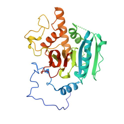
    3ZGF, 3ZGG

  • PubMed Abstract: 

    Glycosyltransferases (GTs) catalyse the sequential addition of monosaccharides to specific acceptor molecules and play major roles in key biological processes. GTs are classified into two main families depending on the inverted or retained stereochemistry of the glycosidic bond formed during the reaction. While the mechanism of inverting enzymes is well characterized, the precise nature of retaining GTs is still a matter of much debate. In an attempt to clarify this issue, studies were initiated to identify reaction-intermediate states by using a crystallographic approach based on caged substrates. In this paper, two distinct structures of AA(Gly)B, a dual-specificity blood group synthase, are described in complex with a UDP-galactose derivative in which the O6'' atom is protected by a 2-nitrobenzyl group. The distinct conformations of the caged substrate in both structures of the enzyme illustrate the highly dynamic nature of its active site. An attempt was also made to photolyse the caged compound at low temperature, which unfortunately is not possible without damaging the uracil group as well. These results pave the way for kinetic crystallography experiments aiming at trapping and characterizing reaction-intermediate states in the mechanism of enzymatic glycosyl transfer.


  • Organizational Affiliation

    Department of Microbiology and Infection Control, Statens Serum Institut, 5 Artillerivej, DK-2300 Copenhagen S, Denmark.


Macromolecules
Find similar proteins by:  (by identity cutoff)  |  3D Structure
Entity ID: 1
MoleculeChains Sequence LengthOrganismDetailsImage
HISTO-BLOOD GROUP ABO SYSTEM TRANSFERASE298Homo sapiensMutation(s): 2 
EC: 2.4.1.37 (PDB Primary Data), 2.4.1.40 (PDB Primary Data)
UniProt & NIH Common Fund Data Resources
Find proteins for P16442 (Homo sapiens)
Explore P16442 
Go to UniProtKB:  P16442
PHAROS:  P16442
Entity Groups  
Sequence Clusters30% Identity50% Identity70% Identity90% Identity95% Identity100% Identity
UniProt GroupP16442
Sequence Annotations
Expand
  • Reference Sequence
Experimental Data & Validation

Experimental Data

  • Method: X-RAY DIFFRACTION
  • Resolution: 1.90 Å
  • R-Value Free: 0.226 
  • R-Value Work: 0.184 
  • R-Value Observed: 0.186 
  • Space Group: C 2 2 21
Unit Cell:
Length ( Å )Angle ( ˚ )
a = 52.687α = 90
b = 150.73β = 90
c = 79.359γ = 90
Software Package:
Software NamePurpose
REFMACrefinement
XDSdata reduction
SCALAdata scaling
PHASERphasing

Structure Validation

View Full Validation Report


Ligand Structure Quality Assessment 


Entry History 

Deposition Data

Revision History  (Full details and data files)

  • Version 1.0: 2014-01-15
    Type: Initial release
  • Version 1.1: 2014-08-06
    Changes: Database references
  • Version 1.2: 2014-08-13
    Changes: Database references
  • Version 1.3: 2023-12-20
    Changes: Data collection, Database references, Derived calculations, Other, Refinement description