4A6N

STRUCTURE OF THE TETRACYCLINE DEGRADING MONOOXYGENASE TETX IN COMPLEX WITH TIGECYCLINE


Experimental Data Snapshot

  • Method: X-RAY DIFFRACTION
  • Resolution: 2.30 Å
  • R-Value Free: 0.260 
  • R-Value Work: 0.218 
  • R-Value Observed: 0.220 

wwPDB Validation   3D Report Full Report


Ligand Structure Quality Assessment 


This is version 1.3 of the entry. See complete history


Literature

Putative Dioxygen-Binding Sites and Recognition of Tigecycline and Minocycline in the Tetracycline-Degrading Monooxygenase Tetx

Volkers, G.Damas, J.M.Palm, G.J.Panjikar, S.Soares, C.M.Hinrichs, W.

(2013) Acta Crystallogr D Biol Crystallogr 69: 1758

  • DOI: https://doi.org/10.1107/S0907444913013802
  • Primary Citation of Related Structures:  
    4A6N, 4A99, 4GUV

  • PubMed Abstract: 

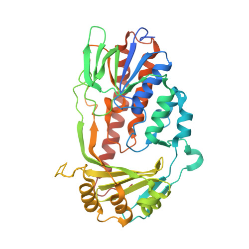
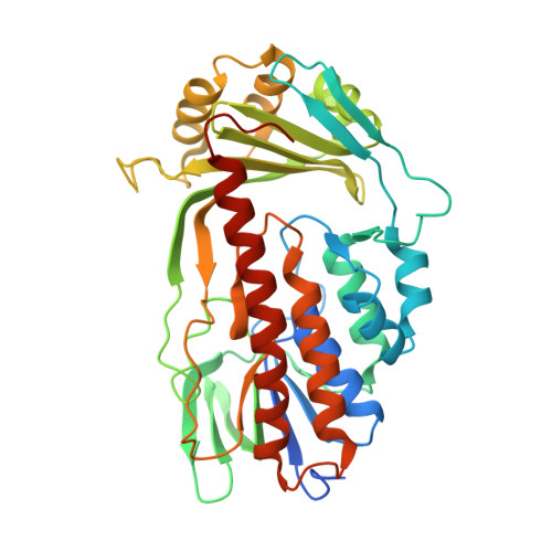
    Expression of the aromatic hydroxylase TetX under aerobic conditions confers bacterial resistance against tetracycline antibiotics. Hydroxylation inactivates and degrades tetracyclines, preventing inhibition of the prokaryotic ribosome. X-ray crystal structure analyses of TetX in complex with the second-generation and third-generation tetracyclines minocycline and tigecycline at 2.18 and 2.30 Å resolution, respectively, explain why both clinically potent antibiotics are suitable substrates. Both tetracyclines bind in a large tunnel-shaped active site in close contact to the cofactor FAD, pre-oriented for regioselective hydroxylation to 11a-hydroxytetracyclines. The characteristic bulky 9-tert-butylglycylamido substituent of tigecycline is solvent-exposed and does not interfere with TetX binding. In the TetX-minocycline complex a second binding site for a minocycline dimer is observed close to the active-site entrance. The pocket is formed by the crystal packing arrangement on the surface of two neighbouring TetX monomers. Crystal structure analysis at 2.73 Å resolution of xenon-pressurized TetX identified two adjacent Xe-binding sites. These putative dioxygen-binding cavities are located in the substrate-binding domain next to the active site. Molecular-dynamics simulations were performed in order to characterize dioxygen-diffusion pathways to FADH2 at the active site.


  • Organizational Affiliation

    Department of Molecular Structural Biology, Institute for Biochemistry, University of Greifswald, Felix-Hausdorff-Strasse 4, Greifswald, Germany.


Macromolecules
Find similar proteins by:  (by identity cutoff)  |  3D Structure
Entity ID: 1
MoleculeChains Sequence LengthOrganismDetailsImage
TETX2 PROTEIN398Bacteroides thetaiotaomicronMutation(s): 0 
UniProt
Find proteins for Q93L51 (Bacteroides thetaiotaomicron)
Explore Q93L51 
Go to UniProtKB:  Q93L51
Entity Groups  
Sequence Clusters30% Identity50% Identity70% Identity90% Identity95% Identity100% Identity
UniProt GroupQ93L51
Sequence Annotations
Expand
  • Reference Sequence
Find similar proteins by:  (by identity cutoff)  |  3D Structure
Entity ID: 2
MoleculeChains Sequence LengthOrganismDetailsImage
TETX2 PROTEIN
B, C, D
398Bacteroides thetaiotaomicronMutation(s): 0 
UniProt
Find proteins for Q93L51 (Bacteroides thetaiotaomicron)
Explore Q93L51 
Go to UniProtKB:  Q93L51
Entity Groups  
Sequence Clusters30% Identity50% Identity70% Identity90% Identity95% Identity100% Identity
UniProt GroupQ93L51
Sequence Annotations
Expand
  • Reference Sequence
Small Molecules
Ligands 3 Unique
IDChains Name / Formula / InChI Key2D Diagram3D Interactions
FAD
Query on FAD

Download Ideal Coordinates CCD File 
E [auth A],
I [auth B],
O [auth C],
S [auth D]
FLAVIN-ADENINE DINUCLEOTIDE
C27 H33 N9 O15 P2
VWWQXMAJTJZDQX-UYBVJOGSSA-N
T1C
Query on T1C

Download Ideal Coordinates CCD File 
F [auth A],
J [auth B],
P [auth C],
T [auth D]
TIGECYCLINE
C29 H41 N5 O8
FPZLLRFZJZRHSY-HJYUBDRYSA-P
SO4
Query on SO4

Download Ideal Coordinates CCD File 
G [auth A]
H [auth A]
K [auth B]
L [auth B]
M [auth B]
G [auth A],
H [auth A],
K [auth B],
L [auth B],
M [auth B],
N [auth B],
Q [auth C],
R [auth C],
U [auth D],
V [auth D],
W [auth D]
SULFATE ION
O4 S
QAOWNCQODCNURD-UHFFFAOYSA-L
Experimental Data & Validation

Experimental Data

  • Method: X-RAY DIFFRACTION
  • Resolution: 2.30 Å
  • R-Value Free: 0.260 
  • R-Value Work: 0.218 
  • R-Value Observed: 0.220 
  • Space Group: P 1
Unit Cell:
Length ( Å )Angle ( ˚ )
a = 68.88α = 110.84
b = 80.79β = 89.84
c = 87.65γ = 93.63
Software Package:
Software NamePurpose
REFMACrefinement
XDSdata reduction
SCALAdata scaling
PHASERphasing

Structure Validation

View Full Validation Report


Ligand Structure Quality Assessment 


Entry History 

Deposition Data

Revision History  (Full details and data files)

  • Version 1.0: 2012-11-21
    Type: Initial release
  • Version 1.1: 2013-08-28
    Changes: Database references
  • Version 1.2: 2013-09-11
    Changes: Database references
  • Version 1.3: 2023-12-20
    Changes: Data collection, Database references, Derived calculations, Other, Refinement description