4GEE

Pyrrolopyrimidine inhibitors of DNA gyrase B and topoisomerase IV, part I: structure guided discovery and optimization of dual targeting agents with potent, broad-spectrum enzymatic activity.


Experimental Data Snapshot

  • Method: X-RAY DIFFRACTION
  • Resolution: 1.70 Å
  • R-Value Free: 0.215 
  • R-Value Work: 0.171 
  • R-Value Observed: 0.174 

wwPDB Validation   3D Report Full Report


Ligand Structure Quality Assessment 


This is version 1.2 of the entry. See complete history


Literature

Pyrrolopyrimidine inhibitors of DNA gyrase B (GyrB) and topoisomerase IV (ParE). Part I: Structure guided discovery and optimization of dual targeting agents with potent, broad-spectrum enzymatic activity.

Tari, L.W.Trzoss, M.Bensen, D.C.Li, X.Chen, Z.Lam, T.Zhang, J.Creighton, C.J.Cunningham, M.L.Kwan, B.Stidham, M.Shaw, K.J.Lightstone, F.C.Wong, S.E.Nguyen, T.B.Nix, J.Finn, J.

(2013) Bioorg Med Chem Lett 23: 1529-1536

  • DOI: https://doi.org/10.1016/j.bmcl.2012.11.032
  • Primary Citation of Related Structures:  
    4GEE, 4GFN, 4GGL, 4HXW, 4HXZ, 4HY1, 4HYM, 4HYP, 4HZ0, 4HZ5

  • PubMed Abstract: 

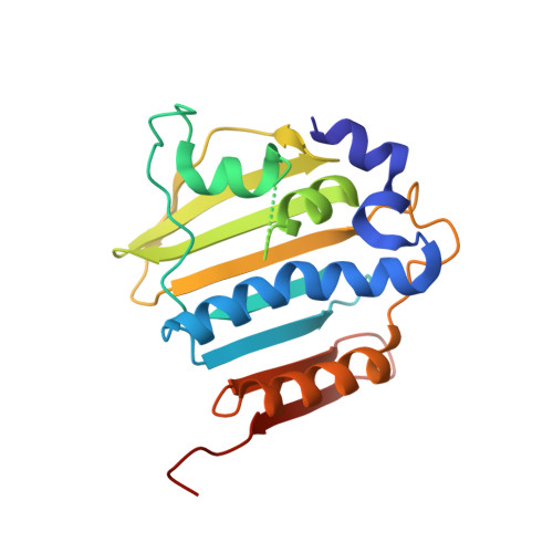
    The bacterial topoisomerases DNA gyrase (GyrB) and topoisomerase IV (ParE) are essential enzymes that control the topological state of DNA during replication. The high degree of conservation in the ATP-binding pockets of these enzymes make them appealing targets for broad-spectrum inhibitor development. A pyrrolopyrimidine scaffold was identified from a pharmacophore-based fragment screen with optimization potential. Structural characterization of inhibitor complexes conducted using selected GyrB/ParE orthologs aided in the identification of important steric, dynamic and compositional differences in the ATP-binding pockets of the targets, enabling the design of highly potent pyrrolopyrimidine inhibitors with broad enzymatic spectrum and dual targeting activity.


  • Organizational Affiliation

    Trius Therapeutics, 6310 Nancy Ridge Dr., San Diego, CA 92121, USA. ltari@triusrx.com


Macromolecules
Find similar proteins by:  (by identity cutoff)  |  3D Structure
Entity ID: 1
MoleculeChains Sequence LengthOrganismDetailsImage
DNA gyrase subunit B215Enterococcus faecalis V583Mutation(s): 0 
Gene Names: gyrBEF_0005
EC: 5.99.1.3
UniProt
Find proteins for Q839Z1 (Enterococcus faecalis (strain ATCC 700802 / V583))
Explore Q839Z1 
Go to UniProtKB:  Q839Z1
Entity Groups  
Sequence Clusters30% Identity50% Identity70% Identity90% Identity95% Identity100% Identity
UniProt GroupQ839Z1
Sequence Annotations
Expand
  • Reference Sequence
Small Molecules
Ligands 2 Unique
IDChains Name / Formula / InChI Key2D Diagram3D Interactions
0WT
Query on 0WT

Download Ideal Coordinates CCD File 
B [auth A](1R,5S,6s)-3-[5-chloro-6-ethyl-2-(pyrimidin-5-yloxy)-7H-pyrrolo[2,3-d]pyrimidin-4-yl]-3-azabicyclo[3.1.0]hexan-6-amine
C17 H18 Cl N7 O
YZRGALHCPBRTES-MSRIBSCDSA-N
GOL
Query on GOL

Download Ideal Coordinates CCD File 
C [auth A]GLYCEROL
C3 H8 O3
PEDCQBHIVMGVHV-UHFFFAOYSA-N
Binding Affinity Annotations 
IDSourceBinding Affinity
0WT PDBBind:  4GEE Ki: 1 (nM) from 1 assay(s)
Binding MOAD:  4GEE Ki: 1 (nM) from 1 assay(s)
Experimental Data & Validation

Experimental Data

  • Method: X-RAY DIFFRACTION
  • Resolution: 1.70 Å
  • R-Value Free: 0.215 
  • R-Value Work: 0.171 
  • R-Value Observed: 0.174 
  • Space Group: P 21 21 21
Unit Cell:
Length ( Å )Angle ( ˚ )
a = 54.93α = 90
b = 58.08β = 90
c = 66.25γ = 90
Software Package:
Software NamePurpose
REFMACrefinement

Structure Validation

View Full Validation Report


Ligand Structure Quality Assessment 


Entry History 

Deposition Data

Revision History  (Full details and data files)

  • Version 1.0: 2013-02-13
    Type: Initial release
  • Version 1.1: 2013-02-27
    Changes: Database references
  • Version 1.2: 2024-02-28
    Changes: Data collection, Database references, Derived calculations