4H2H

Crystal structure of an enolase (mandalate racemase subgroup, target EFI-502101) from Pelagibaca bermudensis htcc2601, with bound mg and l-4-hydroxyproline betaine (betonicine)


Experimental Data Snapshot

  • Method: X-RAY DIFFRACTION
  • Resolution: 1.70 Å
  • R-Value Free: 0.192 
  • R-Value Work: 0.159 
  • R-Value Observed: 0.160 

wwPDB Validation   3D Report Full Report


Ligand Structure Quality Assessment 


This is version 1.3 of the entry. See complete history


Literature

Discovery of new enzymes and metabolic pathways by using structure and genome context.

Zhao, S.Kumar, R.Sakai, A.Vetting, M.W.Wood, B.M.Brown, S.Bonanno, J.B.Hillerich, B.S.Seidel, R.D.Babbitt, P.C.Almo, S.C.Sweedler, J.V.Gerlt, J.A.Cronan, J.E.Jacobson, M.P.

(2013) Nature 502: 698-702

  • DOI: https://doi.org/10.1038/nature12576
  • Primary Citation of Related Structures:  
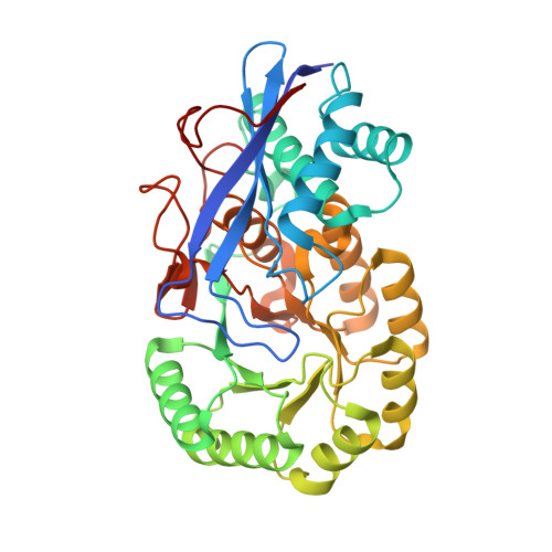
    2PMQ, 4H2H

  • PubMed Abstract: 

    Assigning valid functions to proteins identified in genome projects is challenging: overprediction and database annotation errors are the principal concerns. We and others are developing computation-guided strategies for functional discovery with 'metabolite docking' to experimentally derived or homology-based three-dimensional structures. Bacterial metabolic pathways often are encoded by 'genome neighbourhoods' (gene clusters and/or operons), which can provide important clues for functional assignment. We recently demonstrated the synergy of docking and pathway context by 'predicting' the intermediates in the glycolytic pathway in Escherichia coli. Metabolite docking to multiple binding proteins and enzymes in the same pathway increases the reliability of in silico predictions of substrate specificities because the pathway intermediates are structurally similar. Here we report that structure-guided approaches for predicting the substrate specificities of several enzymes encoded by a bacterial gene cluster allowed the correct prediction of the in vitro activity of a structurally characterized enzyme of unknown function (PDB 2PMQ), 2-epimerization of trans-4-hydroxy-L-proline betaine (tHyp-B) and cis-4-hydroxy-D-proline betaine (cHyp-B), and also the correct identification of the catabolic pathway in which Hyp-B 2-epimerase participates. The substrate-liganded pose predicted by virtual library screening (docking) was confirmed experimentally. The enzymatic activities in the predicted pathway were confirmed by in vitro assays and genetic analyses; the intermediates were identified by metabolomics; and repression of the genes encoding the pathway by high salt concentrations was established by transcriptomics, confirming the osmolyte role of tHyp-B. This study establishes the utility of structure-guided functional predictions to enable the discovery of new metabolic pathways.


  • Organizational Affiliation

    1] Department of Pharmaceutical Chemistry, University of California, San Francisco, California 94143, USA [2].


Macromolecules
Find similar proteins by:  (by identity cutoff)  |  3D Structure
Entity ID: 1
MoleculeChains Sequence LengthOrganismDetailsImage
Mandelate racemase/muconate lactonizing enzyme
A, B, C, D, E
A, B, C, D, E, F, G, H
376Salipiger bermudensis HTCC2601Mutation(s): 0 
Gene Names: R2601_01638
UniProt
Find proteins for Q0FPQ4 (Salipiger bermudensis (strain DSM 26914 / JCM 13377 / KCTC 12554 / HTCC2601))
Explore Q0FPQ4 
Go to UniProtKB:  Q0FPQ4
Entity Groups  
Sequence Clusters30% Identity50% Identity70% Identity90% Identity95% Identity100% Identity
UniProt GroupQ0FPQ4
Sequence Annotations
Expand
  • Reference Sequence
Small Molecules
Ligands 5 Unique
IDChains Name / Formula / InChI Key2D Diagram3D Interactions
0XW
Query on 0XW

Download Ideal Coordinates CCD File 
BB [auth E]
DA [auth C]
FC [auth H]
K [auth A]
LB [auth F]
BB [auth E],
DA [auth C],
FC [auth H],
K [auth A],
LB [auth F],
QA [auth D],
V [auth B],
VB [auth G]
(2S,4R)-4-hydroxy-1,1-dimethylpyrrolidinium-2-carboxylate
C7 H13 N O3
MUNWAHDYFVYIKH-RITPCOANSA-N
IOD
Query on IOD

Download Ideal Coordinates CCD File 
AA [auth B]
AC [auth G]
BA [auth B]
BC [auth G]
CC [auth G]
AA [auth B],
AC [auth G],
BA [auth B],
BC [auth G],
CC [auth G],
DB [auth E],
DC [auth G],
EB [auth E],
FA [auth C],
FB [auth E],
GA [auth C],
GB [auth E],
GC [auth H],
HA [auth C],
HB [auth E],
HC [auth H],
IA [auth C],
IB [auth E],
IC [auth H],
JA [auth C],
JB [auth E],
JC [auth H],
KA [auth C],
KC [auth H],
L [auth A],
LA [auth C],
LC [auth H],
M [auth A],
MA [auth C],
MB [auth F],
N [auth A],
NA [auth C],
NB [auth F],
O [auth A],
OA [auth C],
OB [auth F],
P [auth A],
PB [auth F],
Q [auth A],
QB [auth F],
R [auth A],
RB [auth F],
S [auth A],
SA [auth D],
SB [auth F],
TA [auth D],
TB [auth F],
UA [auth D],
VA [auth D],
W [auth B],
WA [auth D],
WB [auth G],
X [auth B],
XA [auth D],
XB [auth G],
Y [auth B],
YA [auth D],
YB [auth G],
Z [auth B],
ZA [auth D],
ZB [auth G]
IODIDE ION
I
XMBWDFGMSWQBCA-UHFFFAOYSA-M
MPD
Query on MPD

Download Ideal Coordinates CCD File 
CB [auth E],
EA [auth C],
RA [auth D]
(4S)-2-METHYL-2,4-PENTANEDIOL
C6 H14 O2
SVTBMSDMJJWYQN-YFKPBYRVSA-N
NI
Query on NI

Download Ideal Coordinates CCD File 
J [auth A],
U [auth B]
NICKEL (II) ION
Ni
VEQPNABPJHWNSG-UHFFFAOYSA-N
MG
Query on MG

Download Ideal Coordinates CCD File 
AB [auth E]
CA [auth C]
EC [auth H]
I [auth A]
KB [auth F]
AB [auth E],
CA [auth C],
EC [auth H],
I [auth A],
KB [auth F],
PA [auth D],
T [auth B],
UB [auth G]
MAGNESIUM ION
Mg
JLVVSXFLKOJNIY-UHFFFAOYSA-N
Experimental Data & Validation

Experimental Data

  • Method: X-RAY DIFFRACTION
  • Resolution: 1.70 Å
  • R-Value Free: 0.192 
  • R-Value Work: 0.159 
  • R-Value Observed: 0.160 
  • Space Group: P 1 21 1
Unit Cell:
Length ( Å )Angle ( ˚ )
a = 91.035α = 90
b = 152.824β = 105.2
c = 113.027γ = 90
Software Package:
Software NamePurpose
SCALAdata scaling
PHENIXrefinement
PDB_EXTRACTdata extraction
MOSFLMdata reduction
PHENIXphasing

Structure Validation

View Full Validation Report


Ligand Structure Quality Assessment 


Entry History 

Deposition Data

Revision History  (Full details and data files)

  • Version 1.0: 2012-10-10
    Type: Initial release
  • Version 1.1: 2013-09-18
    Changes: Database references
  • Version 1.2: 2013-11-06
    Changes: Database references
  • Version 1.3: 2023-09-20
    Changes: Data collection, Database references, Derived calculations, Refinement description, Structure summary