4L17

GluA2-L483Y-A665C ligand-binding domain in complex with the antagonist DNQX


Experimental Data Snapshot

  • Method: X-RAY DIFFRACTION
  • Resolution: 2.80 Å
  • R-Value Free: 0.244 
  • R-Value Work: 0.195 
  • R-Value Observed: 0.198 

wwPDB Validation   3D Report Full Report


Ligand Structure Quality Assessment 


This is version 1.2 of the entry. See complete history


Literature

A conformational intermediate in glutamate receptor activation.

Lau, A.Y.Salazar, H.Blachowicz, L.Ghisi, V.Plested, A.J.Roux, B.

(2013) Neuron 79: 492-503

  • DOI: https://doi.org/10.1016/j.neuron.2013.06.003
  • Primary Citation of Related Structures:  
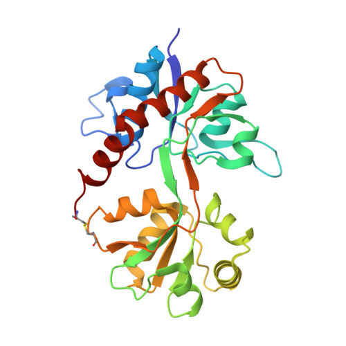
    4L17

  • PubMed Abstract: 

    Ionotropic glutamate receptors (iGluRs) transduce the chemical signal of neurotransmitter release into membrane depolarization at excitatory synapses in the brain. The opening of the transmembrane ion channel of these ligand-gated receptors is driven by conformational transitions that are induced by the association of glutamate molecules to the ligand-binding domains (LBDs). Here, we describe the crystal structure of a GluA2 LBD tetramer in a configuration that involves an ∼30° rotation of the LBD dimers relative to the crystal structure of the full-length receptor. The configuration is stabilized by an engineered disulfide crosslink. Biochemical and electrophysiological studies on full-length receptors incorporating either this crosslink or an engineered metal bridge show that this LBD configuration corresponds to an intermediate state of receptor activation. GluA2 activation therefore involves a combination of both intra-LBD (cleft closure) and inter-LBD dimer conformational transitions. Overall, these results provide a comprehensive structural characterization of an iGluR intermediate state.


  • Organizational Affiliation

    Department of Biophysics and Biophysical Chemistry, Johns Hopkins University School of Medicine, Baltimore, MD 21205, USA. alau@jhmi.edu


Macromolecules
Find similar proteins by:  (by identity cutoff)  |  3D Structure
Entity ID: 1
MoleculeChains Sequence LengthOrganismDetailsImage
Glutamate receptor 2A,
B [auth C],
C [auth E],
D [auth G]
263Rattus norvegicusMutation(s): 2 
Gene Names: Gria2Glur2
UniProt
Find proteins for P19491 (Rattus norvegicus)
Explore P19491 
Go to UniProtKB:  P19491
Entity Groups  
Sequence Clusters30% Identity50% Identity70% Identity90% Identity95% Identity100% Identity
UniProt GroupP19491
Sequence Annotations
Expand
  • Reference Sequence
Experimental Data & Validation

Experimental Data

  • Method: X-RAY DIFFRACTION
  • Resolution: 2.80 Å
  • R-Value Free: 0.244 
  • R-Value Work: 0.195 
  • R-Value Observed: 0.198 
  • Space Group: P 2 21 21
Unit Cell:
Length ( Å )Angle ( ˚ )
a = 48.85α = 90
b = 96.79β = 90
c = 252.84γ = 90
Software Package:
Software NamePurpose
HKL-2000data collection
PHASERphasing
PHENIXrefinement
MOSFLMdata reduction
SCALAdata scaling

Structure Validation

View Full Validation Report


Ligand Structure Quality Assessment 


Entry History 

Deposition Data

Revision History  (Full details and data files)

  • Version 1.0: 2013-08-14
    Type: Initial release
  • Version 1.1: 2013-08-21
    Changes: Database references
  • Version 1.2: 2017-08-02
    Changes: Refinement description, Source and taxonomy