2EFU

The crystal structure of D-amino acid amidase from Ochrobactrum anthropi SV3 complexed with L-phenylalanine


Experimental Data Snapshot

  • Method: X-RAY DIFFRACTION
  • Resolution: 2.30 Å
  • R-Value Free: 0.235 
  • R-Value Work: 0.170 
  • R-Value Observed: 0.174 

wwPDB Validation   3D Report Full Report


Ligand Structure Quality Assessment 


This is version 1.3 of the entry. See complete history


Literature

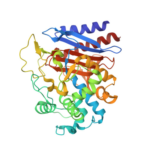
Structures of D-amino-acid amidase complexed with L-phenylalanine and with L-phenylalanine amide: insight into the D-stereospecificity of D-amino-acid amidase from Ochrobactrum anthropi SV3.

Okazaki, S.Suzuki, A.Mizushima, T.Komeda, H.Asano, Y.Yamane, T.

(2008) Acta Crystallogr D Biol Crystallogr 64: 331-334

  • DOI: https://doi.org/10.1107/S0907444907067479
  • Primary Citation of Related Structures:  
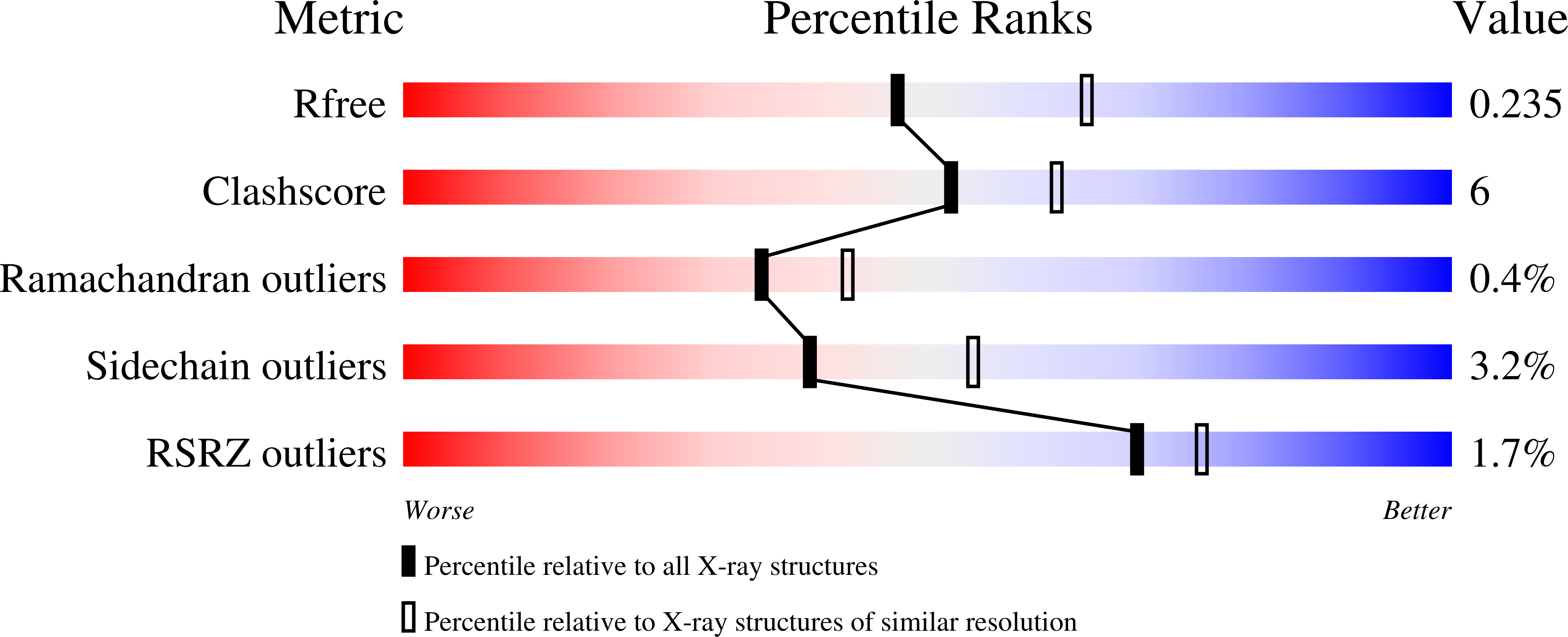
    2EFU, 2EFX

  • PubMed Abstract: 

    The crystal structures of D-amino-acid amidase (DAA) from Ochrobactrum anthropi SV3 in complex with L-phenylalanine and with L-phenylalanine amide were determined at 2.3 and 2.2 A resolution, respectively. Comparison of the L-phenylalanine amide complex with the D-phenylalanine complex reveals that the D-stereospecificity of DAA might be achieved as a consequence of three structural factors: (i) the hydrophobic cavity in the region in which the hydrophobic side chain of the substrate is held, (ii) the spatial arrangement of Gln310 O and Glu114 O epsilon2 that fixes the amino N atom of the substrate and (iii) the existence of two cavities that keep the carboxyl/amide group of the substrate near or apart from Ser60 O gamma.


  • Organizational Affiliation

    Department of Biotechnology, School of Engineering, Nagoya University, Chikusa, Nagoya 464-8603, Japan.


Macromolecules
Find similar proteins by:  (by identity cutoff)  |  3D Structure
Entity ID: 1
MoleculeChains Sequence LengthOrganismDetailsImage
D-Amino acid amidase
A, B, C, D, E
A, B, C, D, E, F
363Brucella anthropiMutation(s): 0 
UniProt
Find proteins for Q9LCC8 (Brucella anthropi)
Explore Q9LCC8 
Go to UniProtKB:  Q9LCC8
Entity Groups  
Sequence Clusters30% Identity50% Identity70% Identity90% Identity95% Identity100% Identity
UniProt GroupQ9LCC8
Sequence Annotations
Expand
  • Reference Sequence
Small Molecules
Ligands 2 Unique
IDChains Name / Formula / InChI Key2D Diagram3D Interactions
PHE
Query on PHE

Download Ideal Coordinates CCD File 
DA [auth E]
K [auth A]
KA [auth F]
N [auth B]
S [auth C]
DA [auth E],
K [auth A],
KA [auth F],
N [auth B],
S [auth C],
Y [auth D]
PHENYLALANINE
C9 H11 N O2
COLNVLDHVKWLRT-QMMMGPOBSA-N
BA
Query on BA

Download Ideal Coordinates CCD File 
AA [auth E]
BA [auth E]
CA [auth E]
EA [auth F]
FA [auth F]
AA [auth E],
BA [auth E],
CA [auth E],
EA [auth F],
FA [auth F],
G [auth A],
GA [auth F],
H [auth A],
HA [auth F],
I [auth A],
IA [auth F],
J [auth A],
JA [auth F],
L [auth B],
M [auth B],
O [auth C],
P [auth C],
Q [auth C],
R [auth C],
T [auth D],
U [auth D],
V [auth D],
W [auth D],
X [auth D],
Z [auth E]
BARIUM ION
Ba
XDFCIPNJCBUZJN-UHFFFAOYSA-N
Experimental Data & Validation

Experimental Data

  • Method: X-RAY DIFFRACTION
  • Resolution: 2.30 Å
  • R-Value Free: 0.235 
  • R-Value Work: 0.170 
  • R-Value Observed: 0.174 
  • Space Group: P 1 21 1
Unit Cell:
Length ( Å )Angle ( ˚ )
a = 77.468α = 90
b = 123.274β = 104.05
c = 116.156γ = 90
Software Package:
Software NamePurpose
REFMACrefinement
JUPITORdata collection
HKL-2000data reduction
SCALEPACKdata scaling
MOLREPphasing

Structure Validation

View Full Validation Report


Ligand Structure Quality Assessment 


Entry History 

Deposition Data

Revision History  (Full details and data files)

  • Version 1.0: 2007-03-06
    Type: Initial release
  • Version 1.1: 2008-02-25
    Changes: Version format compliance
  • Version 1.2: 2011-07-13
    Changes: Version format compliance
  • Version 1.3: 2023-10-25
    Changes: Data collection, Database references, Derived calculations, Refinement description