4GLE

SacUVDE in complex with 6-4PP-containing DNA


Experimental Data Snapshot

  • Method: X-RAY DIFFRACTION
  • Resolution: 2.70 Å
  • R-Value Free: 0.262 
  • R-Value Work: 0.191 
  • R-Value Observed: 0.195 

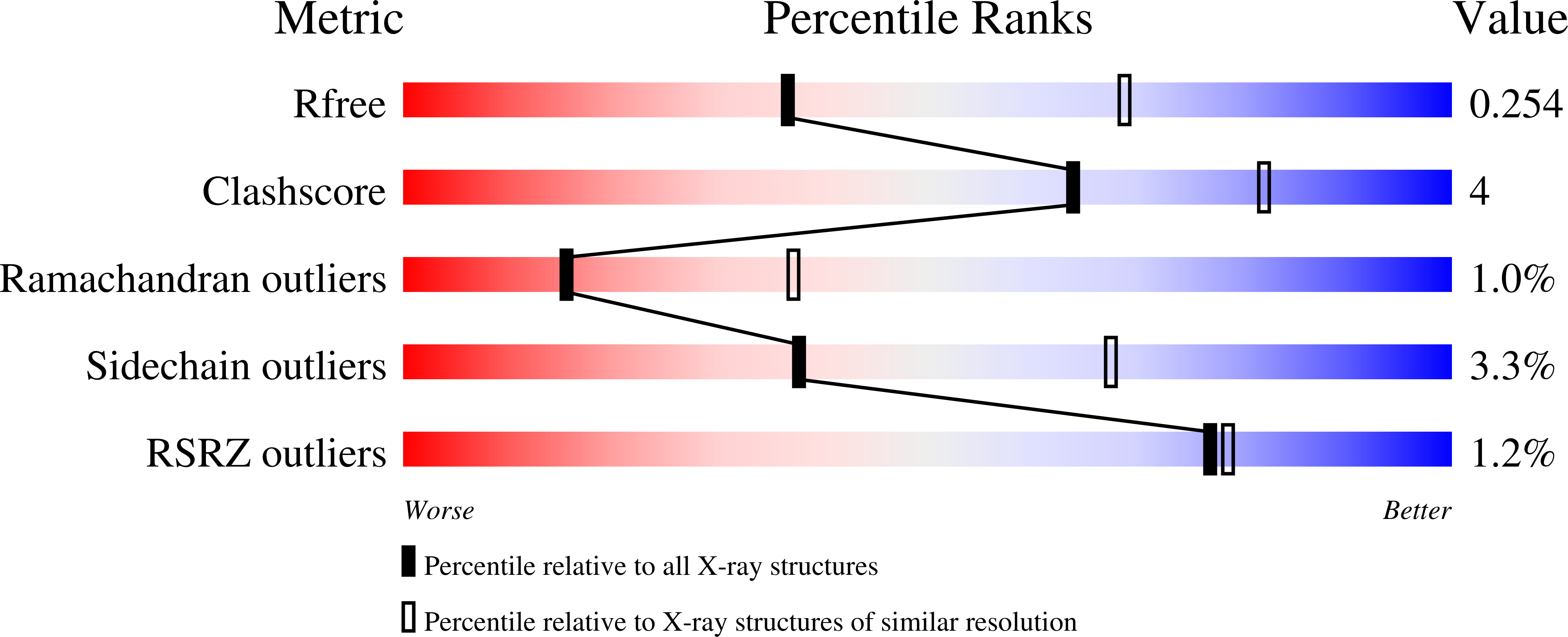
wwPDB Validation   3D Report Full Report


This is version 1.3 of the entry. See complete history


Literature

UV damage endonuclease employs a novel dual-dinucleotide flipping mechanism to recognize different DNA lesions.

Meulenbroek, E.M.Peron Cane, C.Jala, I.Iwai, S.Moolenaar, G.F.Goosen, N.Pannu, N.S.

(2013) Nucleic Acids Res 41: 1363-1371

  • DOI: https://doi.org/10.1093/nar/gks1127
  • Primary Citation of Related Structures:  
    3TC3, 4GLE

  • PubMed Abstract: 

    Repairing damaged DNA is essential for an organism's survival. UV damage endonuclease (UVDE) is a DNA-repair enzyme that can recognize and incise different types of damaged DNA. We present the structure of Sulfolobus acidocaldarius UVDE on its own and in a pre-catalytic complex with UV-damaged DNA containing a 6-4 photoproduct showing a novel 'dual dinucleotide flip' mechanism for recognition of damaged dipyrimidines: the two purines opposite to the damaged pyrimidine bases are flipped into a dipurine-specific pocket, while the damaged bases are also flipped into another cleft.


  • Organizational Affiliation

    Department of Biophysical Structural Chemistry, Leiden Institute of Chemistry, Leiden University, Einsteinweg 55, 2333 CC Leiden, The Netherlands.


Macromolecules

Find similar proteins by:  (by identity cutoff)  |  3D Structure
Entity ID: 3
MoleculeChains Sequence LengthOrganismDetailsImage
UV damage endonucleaseC [auth A]310Sulfolobus acidocaldariusMutation(s): 0 
Gene Names: uvdESaci_1096
EC: 3
UniProt
Find proteins for Q4J9T1 (Sulfolobus acidocaldarius (strain ATCC 33909 / DSM 639 / JCM 8929 / NBRC 15157 / NCIMB 11770))
Explore Q4J9T1 
Go to UniProtKB:  Q4J9T1
Entity Groups  
Sequence Clusters30% Identity50% Identity70% Identity90% Identity95% Identity100% Identity
UniProt GroupQ4J9T1
Sequence Annotations
Expand
  • Reference Sequence

Find similar nucleic acids by:  Sequence   |   3D Structure  

Entity ID: 1
MoleculeChains LengthOrganismImage
5'-D(*CP*GP*TP*CP*GP*TP*CP*AP*AP*GP*GP*AP*CP*GP*C)-3'A [auth B]15N/A
Sequence Annotations
Expand
  • Reference Sequence

Find similar nucleic acids by:  Sequence   |   3D Structure  

Entity ID: 2
MoleculeChains LengthOrganismImage
5'-D(*GP*CP*GP*TP*CP*CP*(64T)P*(5PY)P*GP*AP*CP*GP*AP*CP*G)-3'B [auth C]15N/A
Sequence Annotations
Expand
  • Reference Sequence
Experimental Data & Validation

Experimental Data

  • Method: X-RAY DIFFRACTION
  • Resolution: 2.70 Å
  • R-Value Free: 0.262 
  • R-Value Work: 0.191 
  • R-Value Observed: 0.195 
  • Space Group: C 2 2 21
Unit Cell:
Length ( Å )Angle ( ˚ )
a = 57.2α = 90
b = 112.51β = 90
c = 153.85γ = 90
Software Package:
Software NamePurpose
MxCuBEdata collection
MOLREPphasing
REFMACrefinement
iMOSFLMdata reduction
SCALAdata scaling

Structure Validation

View Full Validation Report


Entry History 

Deposition Data

Revision History  (Full details and data files)

  • Version 1.0: 2012-11-21
    Type: Initial release
  • Version 1.1: 2012-12-26
    Changes: Database references
  • Version 1.2: 2013-02-06
    Changes: Database references
  • Version 1.3: 2023-09-13
    Changes: Data collection, Database references, Derived calculations, Refinement description