4M30

Crystal structure of RNASE III complexed with double-stranded RNA AND AMP (TYPE II CLEAVAGE)


Experimental Data Snapshot

  • Method: X-RAY DIFFRACTION
  • Resolution: 2.50 Å
  • R-Value Free: 0.261 
  • R-Value Work: 0.198 
  • R-Value Observed: 0.200 

wwPDB Validation   3D Report Full Report


Ligand Structure Quality Assessment 


This is version 1.2 of the entry. See complete history


Literature

RNase III: Genetics and Function; Structure and Mechanism.

Court, D.L.Gan, J.Liang, Y.H.Shaw, G.X.Tropea, J.E.Costantino, N.Waugh, D.S.Ji, X.

(2013) Annu Rev Genet 47: 405-431

  • DOI: https://doi.org/10.1146/annurev-genet-110711-155618
  • Primary Citation of Related Structures:  
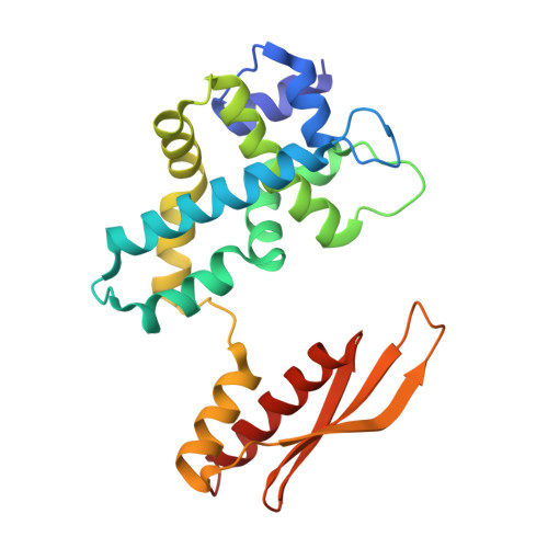
    4M2Z, 4M30

  • PubMed Abstract: 

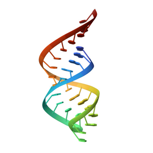
    RNase III is a global regulator of gene expression in Escherichia coli that is instrumental in the maturation of ribosomal and other structural RNAs. We examine here how RNase III itself is regulated in response to growth and other environmental changes encountered by the cell and how, by binding or processing double-stranded RNA (dsRNA) intermediates, RNase III controls the expression of genes. Recent insight into the mechanism of dsRNA binding and processing, gained from structural studies of RNase III, is reviewed. Structural studies also reveal new cleavage sites in the enzyme that can generate longer 3' overhangs.


  • Organizational Affiliation

    Center for Cancer Research, National Cancer Institute, Frederick, Maryland 21702; email: courtd@mail.nih.gov , ganj@mail.nih.gov , liangyh@mail.nih.gov , shawg@mail.nih.gov , costantn@mail.nih.gov , tropeajo@mail.nih.gov , waughd@mail.nih.gov , jix@mail.nih.gov.


Macromolecules

Find similar proteins by:  (by identity cutoff)  |  3D Structure
Entity ID: 1
MoleculeChains Sequence LengthOrganismDetailsImage
Ribonuclease 3
A, B
221Aquifex aeolicus VF5Mutation(s): 0 
Gene Names: aq_946rnc
EC: 3.1.26.3
UniProt
Find proteins for O67082 (Aquifex aeolicus (strain VF5))
Explore O67082 
Go to UniProtKB:  O67082
Entity Groups  
Sequence Clusters30% Identity50% Identity70% Identity90% Identity95% Identity100% Identity
UniProt GroupO67082
Sequence Annotations
Expand
  • Reference Sequence
Find similar nucleic acids by:  (by identity cutoff)  |  3D Structure
Entity ID: 2
MoleculeChains LengthOrganismImage
RNA12
C, D
27N/A
Sequence Annotations
Expand
  • Reference Sequence
Small Molecules
Ligands 3 Unique
IDChains Name / Formula / InChI Key2D Diagram3D Interactions
A
Query on A

Download Ideal Coordinates CCD File 
L [auth C],
P [auth D]
ADENOSINE-5'-MONOPHOSPHATE
C10 H14 N5 O7 P
UDMBCSSLTHHNCD-KQYNXXCUSA-N
MPD
Query on MPD

Download Ideal Coordinates CCD File 
I [auth B](4S)-2-METHYL-2,4-PENTANEDIOL
C6 H14 O2
SVTBMSDMJJWYQN-YFKPBYRVSA-N
MG
Query on MG

Download Ideal Coordinates CCD File 
E [auth A]
F [auth A]
G [auth B]
H [auth B]
J [auth C]
E [auth A],
F [auth A],
G [auth B],
H [auth B],
J [auth C],
K [auth C],
M [auth D],
N [auth D],
O [auth D]
MAGNESIUM ION
Mg
JLVVSXFLKOJNIY-UHFFFAOYSA-N
Experimental Data & Validation

Experimental Data

  • Method: X-RAY DIFFRACTION
  • Resolution: 2.50 Å
  • R-Value Free: 0.261 
  • R-Value Work: 0.198 
  • R-Value Observed: 0.200 
  • Space Group: P 32 2 1
Unit Cell:
Length ( Å )Angle ( ˚ )
a = 81.104α = 90
b = 81.104β = 90
c = 223.962γ = 120
Software Package:
Software NamePurpose
SCALEPACKdata scaling
PHENIXrefinement
PDB_EXTRACTdata extraction
SERGUIdata collection
HKL-2000data reduction
PHASERphasing

Structure Validation

View Full Validation Report


Ligand Structure Quality Assessment 


Entry History 

Deposition Data

Revision History  (Full details and data files)

  • Version 1.0: 2013-12-11
    Type: Initial release
  • Version 1.1: 2023-08-30
    Changes: Data collection, Database references, Derived calculations, Structure summary
  • Version 1.2: 2023-09-20
    Changes: Refinement description