5TDZ

TEV Cleaved Human ATP Citrate Lyase Bound to Tartrate and ADP


Experimental Data Snapshot

  • Method: X-RAY DIFFRACTION
  • Resolution: 2.00 Å
  • R-Value Free: 0.222 
  • R-Value Work: 0.175 
  • R-Value Observed: 0.177 

wwPDB Validation   3D Report Full Report


Ligand Structure Quality Assessment 


This is version 1.1 of the entry. See complete history


Literature

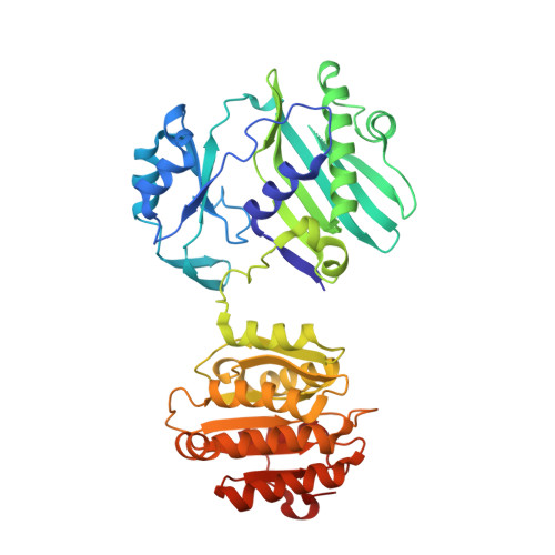
Binding of hydroxycitrate to human ATP-citrate lyase.

Hu, J.Komakula, A.Fraser, M.E.

(2017) Acta Crystallogr D Struct Biol 73: 660-671

  • DOI: https://doi.org/10.1107/S2059798317009871
  • Primary Citation of Related Structures:  
    5TDE, 5TDF, 5TDM, 5TDZ, 5TE1, 5TEQ, 5TES, 5TET

  • PubMed Abstract: 

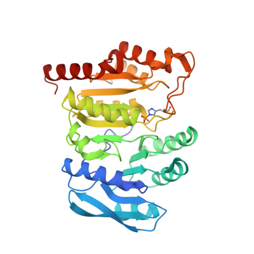
    Hydroxycitrate from the fruit of Garcinia cambogia [i.e. (2S,3S)-2-hydroxycitrate] is the best-known inhibitor of ATP-citrate lyase. Well diffracting crystals showing how the inhibitor binds to human ATP-citrate lyase were grown by modifying the protein. The protein was modified by introducing cleavage sites for Tobacco etch virus protease on either side of a disordered linker. The protein crystallized consisted of residues 2-425-ENLYFQ and S-488-810 of human ATP-citrate lyase. (2S,3S)-2-Hydroxycitrate binds in the same orientation as citrate, but the citrate-binding domain (residues 248-421) adopts a different orientation with respect to the rest of the protein (residues 4-247, 490-746 and 748-809) from that previously seen. For the first time, electron density was evident for the loop that contains His760, which is phosphorylated as part of the catalytic mechanism. The pro-S carboxylate of (2S,3S)-2-hydroxycitrate is available to accept a phosphoryl group from His760. However, when co-crystals were grown with ATP and magnesium ions as well as either the inhibitor or citrate, Mg 2+ -ADP was bound and His760 was phosphorylated. The phosphoryl group was not transferred to the organic acid. This led to the interpretation that the active site is trapped in an open conformation. The strategy of designing cleavage sites to remove disordered residues could be useful in determining the crystal structures of other proteins.


  • Organizational Affiliation

    Department of Biological Sciences, University of Calgary, 2500 University Drive NW, Calgary, Alberta T2N 1N4, Canada.


Macromolecules
Find similar proteins by:  (by identity cutoff)  |  3D Structure
Entity ID: 1
MoleculeChains Sequence LengthOrganismDetailsImage
ATP-citrate synthase431Homo sapiensMutation(s): 0 
Gene Names: ACLY
EC: 2.3.3.8
UniProt & NIH Common Fund Data Resources
Find proteins for P53396 (Homo sapiens)
Explore P53396 
Go to UniProtKB:  P53396
PHAROS:  P53396
GTEx:  ENSG00000131473 
Entity Groups  
Sequence Clusters30% Identity50% Identity70% Identity90% Identity95% Identity100% Identity
UniProt GroupP53396
Sequence Annotations
Expand
  • Reference Sequence
Find similar proteins by:  (by identity cutoff)  |  3D Structure
Entity ID: 2
MoleculeChains Sequence LengthOrganismDetailsImage
ATP-citrate synthase324Homo sapiensMutation(s): 0 
Gene Names: ACLY
EC: 2.3.3.8
UniProt & NIH Common Fund Data Resources
Find proteins for P53396 (Homo sapiens)
Explore P53396 
Go to UniProtKB:  P53396
PHAROS:  P53396
GTEx:  ENSG00000131473 
Entity Groups  
Sequence Clusters30% Identity50% Identity70% Identity90% Identity95% Identity100% Identity
UniProt GroupP53396
Sequence Annotations
Expand
  • Reference Sequence
Small Molecules
Ligands 8 Unique
IDChains Name / Formula / InChI Key2D Diagram3D Interactions
ADP
Query on ADP

Download Ideal Coordinates CCD File 
D [auth A]ADENOSINE-5'-DIPHOSPHATE
C10 H15 N5 O10 P2
XTWYTFMLZFPYCI-KQYNXXCUSA-N
ADN
Query on ADN

Download Ideal Coordinates CCD File 
G [auth A]ADENOSINE
C10 H13 N5 O4
OIRDTQYFTABQOQ-KQYNXXCUSA-N
PGE
Query on PGE

Download Ideal Coordinates CCD File 
I [auth A],
L [auth B]
TRIETHYLENE GLYCOL
C6 H14 O4
ZIBGPFATKBEMQZ-UHFFFAOYSA-N
TLA
Query on TLA

Download Ideal Coordinates CCD File 
C [auth A]L(+)-TARTARIC ACID
C4 H6 O6
FEWJPZIEWOKRBE-JCYAYHJZSA-N
TRS
Query on TRS

Download Ideal Coordinates CCD File 
J [auth B]2-AMINO-2-HYDROXYMETHYL-PROPANE-1,3-DIOL
C4 H12 N O3
LENZDBCJOHFCAS-UHFFFAOYSA-O
PO4
Query on PO4

Download Ideal Coordinates CCD File 
F [auth A]PHOSPHATE ION
O4 P
NBIIXXVUZAFLBC-UHFFFAOYSA-K
GOL
Query on GOL

Download Ideal Coordinates CCD File 
H [auth A],
K [auth B]
GLYCEROL
C3 H8 O3
PEDCQBHIVMGVHV-UHFFFAOYSA-N
MG
Query on MG

Download Ideal Coordinates CCD File 
E [auth A]MAGNESIUM ION
Mg
JLVVSXFLKOJNIY-UHFFFAOYSA-N
Modified Residues  1 Unique
IDChains TypeFormula2D DiagramParent
NEP
Query on NEP
B
L-PEPTIDE LINKINGC6 H10 N3 O5 PHIS
Binding Affinity Annotations 
IDSourceBinding Affinity
ADP Binding MOAD:  5TDZ Ki: 1.50e+5 (nM) from 1 assay(s)
Experimental Data & Validation

Experimental Data

  • Method: X-RAY DIFFRACTION
  • Resolution: 2.00 Å
  • R-Value Free: 0.222 
  • R-Value Work: 0.175 
  • R-Value Observed: 0.177 
  • Space Group: P 21 21 21
Unit Cell:
Length ( Å )Angle ( ˚ )
a = 55.672α = 90
b = 84.291β = 90
c = 195.594γ = 90
Software Package:
Software NamePurpose
PHENIXrefinement

Structure Validation

View Full Validation Report


Ligand Structure Quality Assessment 


Entry History 

Deposition Data

Revision History  (Full details and data files)

  • Version 1.0: 2017-08-09
    Type: Initial release
  • Version 1.1: 2017-08-16
    Changes: Database references