6TZR

A DNA G-quadruplex/i-motif hybrid

  • Classification: DNA
  • Organism(s): synthetic construct
  • Mutation(s): No 

  • Deposited: 2019-08-13 Released: 2019-10-16 
  • Deposition Author(s): Chu, B., Paukstelis, P.J.
  • Funding Organization(s): National Science Foundation (NSF, United States)

Experimental Data Snapshot

  • Method: X-RAY DIFFRACTION
  • Resolution: 2.40 Å
  • R-Value Free: 0.254 
  • R-Value Work: 0.212 
  • R-Value Observed: 0.218 

wwPDB Validation   3D Report Full Report


This is version 1.3 of the entry. See complete history


Literature

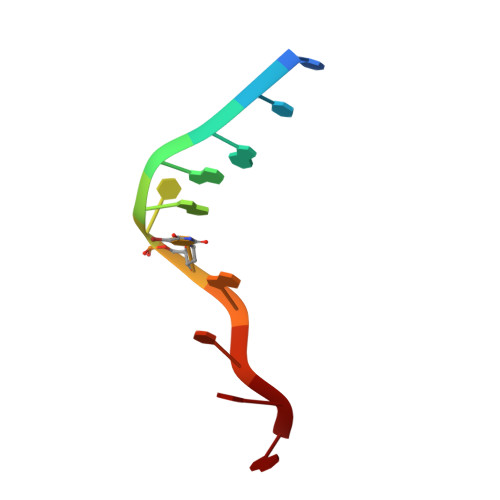
A DNA G-quadruplex/i-motif hybrid.

Chu, B.Zhang, D.Paukstelis, P.J.

(2019) Nucleic Acids Res 47: 11921-11930

  • DOI: https://doi.org/10.1093/nar/gkz1008
  • Primary Citation of Related Structures:  
    6TZQ, 6TZR, 6TZS

  • PubMed Abstract: 

    DNA can form many structures beyond the canonical Watson-Crick double helix. It is now clear that noncanonical structures are present in genomic DNA and have biological functions. G-rich G-quadruplexes and C-rich i-motifs are the most well-characterized noncanonical DNA motifs that have been detected in vivo with either proscribed or postulated biological roles. Because of their independent sequence requirements, these structures have largely been considered distinct types of quadruplexes. Here, we describe the crystal structure of the DNA oligonucleotide, d(CCAGGCTGCAA), that self-associates to form a quadruplex structure containing two central antiparallel G-tetrads and six i-motif C-C+ base pairs. Solution studies suggest a robust structural motif capable of assembling as a tetramer of individual strands or as a dimer when composed of tandem repeats. This hybrid structure highlights the growing structural diversity of DNA and suggests that biological systems may harbor many functionally important non-duplex structures.


  • Organizational Affiliation

    Department of Chemistry and Biochemistry, Center for Biomolecular Structure and Organization, University of Maryland, College Park, MD 20742, USA.


Macromolecules

Find similar nucleic acids by:  Sequence   |   3D Structure  

Entity ID: 1
MoleculeChains LengthOrganismImage
DNA (5'-D(*CP*CP*AP*GP*GP*CP*(BRU)P*GP*CP*AP*A)-3')
A, B
11synthetic construct
Sequence Annotations
Expand
  • Reference Sequence
Small Molecules
Ligands 1 Unique
IDChains Name / Formula / InChI Key2D Diagram3D Interactions
BA
Query on BA

Download Ideal Coordinates CCD File 
C [auth A]BARIUM ION
Ba
XDFCIPNJCBUZJN-UHFFFAOYSA-N
Experimental Data & Validation

Experimental Data

  • Method: X-RAY DIFFRACTION
  • Resolution: 2.40 Å
  • R-Value Free: 0.254 
  • R-Value Work: 0.212 
  • R-Value Observed: 0.218 
  • Space Group: P 32 2 1
Unit Cell:
Length ( Å )Angle ( ˚ )
a = 37.099α = 90
b = 37.099β = 90
c = 98.309γ = 120
Software Package:
Software NamePurpose
Aimlessdata scaling
REFMACrefinement
PDB_EXTRACTdata extraction
XDSdata reduction
SHELXCDphasing

Structure Validation

View Full Validation Report


Entry History & Funding Information

Deposition Data


Funding OrganizationLocationGrant Number
National Science Foundation (NSF, United States)United States1149665

Revision History  (Full details and data files)

  • Version 1.0: 2019-10-16
    Type: Initial release
  • Version 1.1: 2019-11-27
    Changes: Author supporting evidence, Database references
  • Version 1.2: 2019-12-25
    Changes: Database references
  • Version 1.3: 2024-03-13
    Changes: Data collection, Database references, Derived calculations