6AS9

Filamentous Assembly of Green Fluorescent Protein Supported by a C-terminal fusion of 18-residues, viewed in space group P212121 form 2


Experimental Data Snapshot

  • Method: X-RAY DIFFRACTION
  • Resolution: 1.75 Å
  • R-Value Free: 0.183 
  • R-Value Work: 0.156 
  • R-Value Observed: 0.157 

wwPDB Validation   3D Report Full Report


This is version 1.2 of the entry. See complete history


Literature

Atomic insights into the genesis of cellular filaments by globular proteins.

McPartland, L.Heller, D.M.Eisenberg, D.S.Hochschild, A.Sawaya, M.R.

(2018) Nat Struct Mol Biol 25: 705-714

  • DOI: https://doi.org/10.1038/s41594-018-0096-7
  • Primary Citation of Related Structures:  
    6AS9

  • PubMed Abstract: 

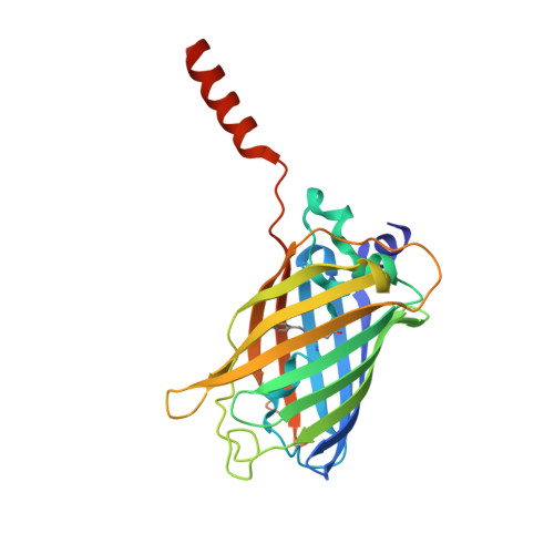
    Self-assembly of proteins into filaments, such as actin and tubulin filaments, underlies essential cellular processes in all three domains of life. The early emergence of filaments in evolutionary history suggests that filament genesis might be a robust process. Here we describe the fortuitous construction of GFP fusion proteins that self-assemble as fluorescent polar filaments in Escherichia coli. Filament formation is achieved by appending as few as 12 residues to GFP. Crystal structures reveal that each protomer donates an appendage to fill a groove between the two following protomers along the filament. This exchange of appendages resembles runaway domain swapping but is distinguished by higher efficiency because monomers cannot competitively bind their own appendages. Ample evidence for this 'runaway domain coupling' mechanism in nature suggests it could facilitate the evolutionary pathway from globular protein to polar filament, requiring a minimal extension of protein sequence and no substantial refolding.


  • Organizational Affiliation

    Department of Microbiology and Immunobiology, Harvard Medical School, Boston, MA, USA.


Macromolecules
Find similar proteins by:  (by identity cutoff)  |  3D Structure
Entity ID: 1
MoleculeChains Sequence LengthOrganismDetailsImage
Green fluorescent protein254Aequorea victoriaMutation(s): 1 
Gene Names: GFP
UniProt
Find proteins for P42212 (Aequorea victoria)
Explore P42212 
Go to UniProtKB:  P42212
Entity Groups  
Sequence Clusters30% Identity50% Identity70% Identity90% Identity95% Identity100% Identity
UniProt GroupP42212
Sequence Annotations
Expand
  • Reference Sequence
Experimental Data & Validation

Experimental Data

  • Method: X-RAY DIFFRACTION
  • Resolution: 1.75 Å
  • R-Value Free: 0.183 
  • R-Value Work: 0.156 
  • R-Value Observed: 0.157 
  • Space Group: P 21 21 21
Unit Cell:
Length ( Å )Angle ( ˚ )
a = 51.1α = 90
b = 54.52β = 90
c = 85.47γ = 90
Software Package:
Software NamePurpose
XSCALEdata scaling
PHASERphasing
BUSTERrefinement
PDB_EXTRACTdata extraction
XDSdata reduction
Cootmodel building

Structure Validation

View Full Validation Report


Entry History & Funding Information

Deposition Data


Funding OrganizationLocationGrant Number
Howard Hughes Medical Institute (HHMI)United States1616265
National Institutes of Health/Office of the DirectorUnited StatesOD003806
National Institutes of Health/National Institute of General Medical Sciences (NIH/NIGMS)United StatesGM115941

Revision History  (Full details and data files)

  • Version 1.0: 2018-05-30
    Type: Initial release
  • Version 1.1: 2018-12-12
    Changes: Data collection, Database references
  • Version 1.2: 2019-11-20
    Changes: Author supporting evidence