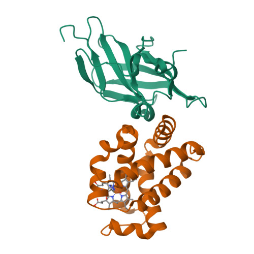
Structure Determination MethodologyScientific Name of Source OrganismRefinement Resolution (Å)Enzyme Classification NameMembrane Protein Annotation | Pilpa, R.M., Fadeev, E.A., Villareal, V.A., Wong, M.A., Phillips, M., Clubb, R.T. (2006) J Mol Biology 360: 435-447 | Released | 2006-08-22 | | Method | SOLUTION NMR | | Organisms | | | Macromolecule | |
Jacques, D.A., Krishna Kumar, K., Caradoc-Davies, T.T., Langley, D.B., Mackay, J.P., Guss, J.M., Gell, D.A. To be published | Released | 2011-09-21 | | Method | X-RAY DIFFRACTION 2.83 Å | | Organisms | | | Macromolecule | | | Unique Ligands | HEM |
Zhu, X., Naismith, J.H. (2012) Acta Crystallogr Sect F Struct Biol Cryst Commun 68: 610 | Released | 2012-02-15 | | Method | X-RAY DIFFRACTION 1.73 Å | | Organisms | | | Macromolecule | | | Unique Ligands | SO4, ZN |
Zhu, X., Naismith, J.H. (2012) Acta Crystallogr Sect F Struct Biol Cryst Commun 68: 610 | Released | 2012-02-15 | | Method | X-RAY DIFFRACTION 2.22 Å | | Organisms | | | Macromolecule | | | Unique Ligands | ADK, SO4, ZN |
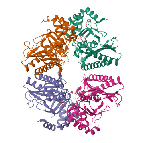
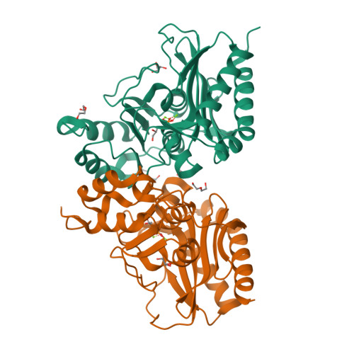
Bhattacharyya, S., Dutta, D., Ghosh, A.K., Das, A.K. (2014) FEBS J 281: 5309-5324 | Released | 2013-07-24 | | Method | X-RAY DIFFRACTION 2.3 Å | | Organisms | | | Macromolecule | | | Unique Ligands | CL, DTO, GOL, MG, P33, PG4, PO4 |
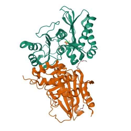
Dutta, A., Bhattacharyya, S., Dutta, D., Das, A.K. (2014) FEBS J 281: 5309-5324 | Released | 2013-11-27 | | Method | X-RAY DIFFRACTION 2.04 Å | | Organisms | | | Macromolecule | | | Unique Ligands | GOL, MG, PG4, PO4 |
Dutta, A., Bhattacharyya, S., Dutta, D., Das, A.K. (2014) FEBS J 281: 5309-5324 | Released | 2013-11-27 | | Method | X-RAY DIFFRACTION 2.5 Å | | Organisms | | | Macromolecule | | | Unique Ligands | GOL, MG, PG4, PO4 |
Minasov, G., Nocadello, S., Shuvalova, L., Shatsman, S., Kwon, K., Bagnoli, F., Falugi, F., Bottomley, M., Grandi, G., Anderson, W.F., Center for Structural Genomics of Infectious Diseases (CSGID) (2016) Acta Crystallogr D Struct Biol 72: 113-120 | Released | 2014-05-07 | | Method | X-RAY DIFFRACTION 1.7 Å | | Organisms | | | Macromolecule | | | Unique Ligands | BTB |
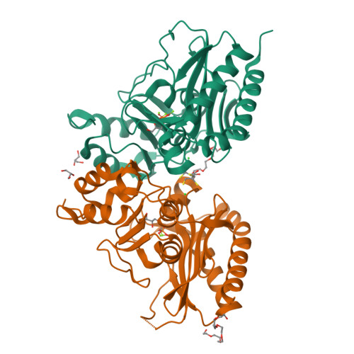
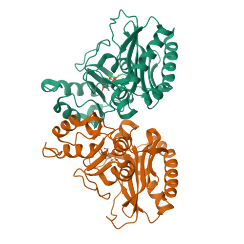
Liu, X., Lan, L., Yang, C.G. (2014) Proc Natl Acad Sci U S A 111: E4981-E4990 | Released | 2014-11-19 | | Method | X-RAY DIFFRACTION 2.21 Å | | Organisms | | | Macromolecule | | | Unique Ligands | CL |
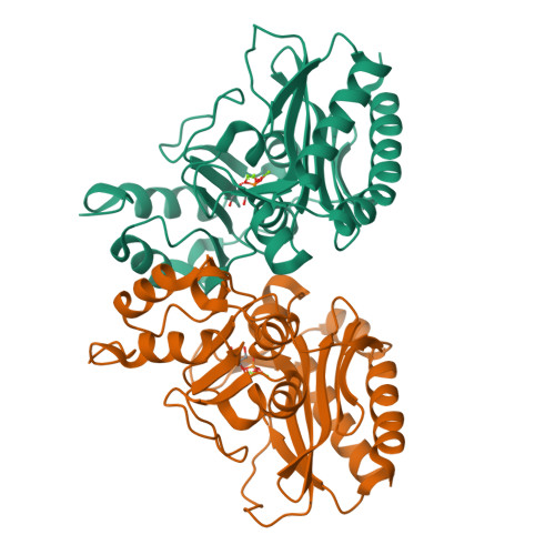
Deng, X. (2016) Nat Commun 7: 13376-13376 | Released | 2016-09-14 | | Method | X-RAY DIFFRACTION 3.17 Å | | Organisms | | | Macromolecule | |
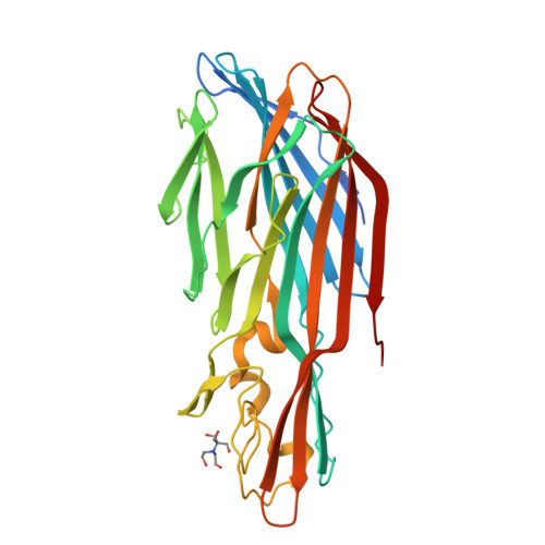
Zhang, S., Wei, J., Wu, S., Zhang, X., Luo, M., Wang, D. To be published | Released | 2017-03-22 | | Method | X-RAY DIFFRACTION 1.25 Å | | Organisms | | | Macromolecule | |
|