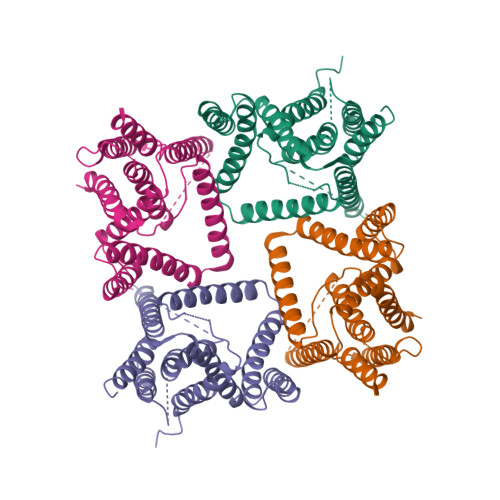
Structure Determination MethodologyScientific Name of Source OrganismMore... Refinement Resolution (Å)Enzyme Classification NameMembrane Protein Annotation | Yao, X., Chong, H.H., Waltersperger, S., Wang, M.T., He, Y.X., Cui, S. (2013) FASEB J 27: 1203-1213 | Released | 2012-12-19 | | Method | X-RAY DIFFRACTION 2.087 Å | | Organisms | | | Macromolecule | | | Unique Ligands | NH4, SO4 |
Yao, X., Chong, H.H., Waltersperger, S., Wang, M.T., He, Y.X., Cui, S. (2013) FASEB J 27: 1203-1213 | Released | 2012-12-19 | | Method | X-RAY DIFFRACTION 2.324 Å | | Organisms | | | Macromolecule | | | Unique Ligands | SO4 |
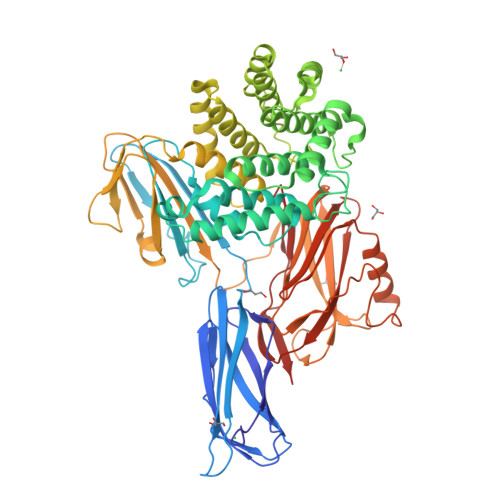
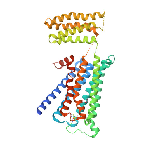
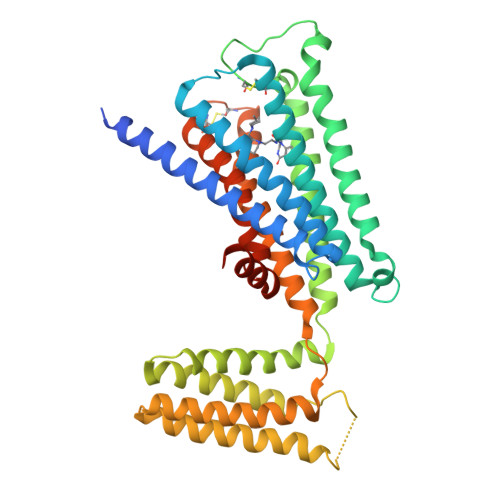
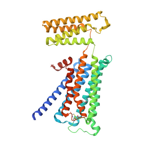
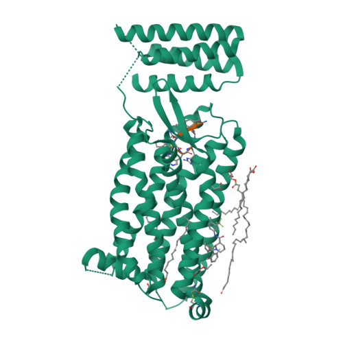
Thorsen, T.S., Matt, R., Weis, W.I., Kobilka, B. (2014) Structure 22: 1657-1664 | Released | 2014-11-26 | | Method | X-RAY DIFFRACTION 2.8 Å | | Organisms | | | Macromolecule | | | Unique Ligands | 0HK, OLC, P6G, TAR |
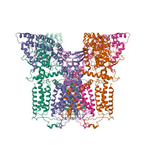
Dang, S., Wu, S., Wang, J., Shi, Y. (2015) Proc Natl Acad Sci U S A 112: 3344-3349 | Released | 2015-03-18 | | Method | X-RAY DIFFRACTION 3.855 Å | | Organisms | | | Macromolecule | | | Unique Ligands | 4B5 |
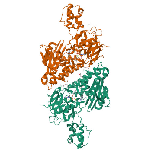
Garcia-Ferrer, I., Arede, P., Gomez-Blanco, J., Luque, D., Duquerroy, S., Caston, J.R., Goulas, T., Gomis-Ruth, X.F. (2015) Proc Natl Acad Sci U S A 112: 8290-8295 | Released | 2015-06-10 | | Method | X-RAY DIFFRACTION 2.7 Å | | Organisms | | | Macromolecule | | | Unique Ligands | GOL, NI |
Yu, Y., Yang, Y.Q., Li, X.L., Yu, J., Ge, J.P., Li, J., Rao, Y., Yang, M.J. (2017) J Med Chem 60: 1994-2005 | Released | 2017-03-22 | | Method | X-RAY DIFFRACTION 2.162 Å | | Organisms | | | Macromolecule | | | Unique Ligands | ACT, FAD, MG, TRT |
Yu, Y., Li, X.L. (2017) J Med Chem 60: 1994-2005 | Released | 2017-03-29 | | Method | X-RAY DIFFRACTION 2.7 Å | | Organisms | | | Macromolecule | | | Unique Ligands | ACT, CXS, FAD, MG, NAD, TRT |
Suno, R., Maeda, S., Yasuda, S., Yamashita, K., Hirata, K., Horita, S., Tawaramoto, M.S., Tsujimoto, H., Murata, T., Kinoshita, M., Yamamoto, M., Kobilka, B.K., Iwata, S., Kobayashi, T. (2018) Nat Chem Biol 14: 1150-1158 | Released | 2018-11-21 | | Method | X-RAY DIFFRACTION 2.6 Å | | Organisms | | | Macromolecule | | | Unique Ligands | QNB |
Suno, R., Maeda, S., Yasuda, S., Yamashita, K., Hirata, K., Horita, S., Tawaramoto, M.S., Tsujimoto, H., Murata, T., Kinoshita, M., Yamamoto, M., Kobilka, B.K., Iwata, S., Kobayashi, T. (2018) Nat Chem Biol 14: 1150-1158 | Released | 2018-11-21 | | Method | X-RAY DIFFRACTION 3 Å | | Organisms | | | Macromolecule | | | Unique Ligands | 3C0 |
Suno, R., Maeda, S., Yasuda, S., Yamashita, K., Hirata, K., Horita, S., Tawaramoto, M.S., Tsujimoto, H., Murata, T., Kinoshita, M., Yamamoto, M., Kobilka, B.K., Iwata, S., Kobayashi, T. (2018) Nat Chem Biol 14: 1150-1158 | Released | 2018-11-21 | | Method | X-RAY DIFFRACTION 2.95 Å | | Organisms | | | Macromolecule | | | Unique Ligands | 82F |
Suno, R., Maeda, S., Yasuda, S., Yamashita, K., Hirata, K., Horita, S., Tawaramoto, M.S., Tsujimoto, H., Murata, T., Kinoshita, M., Yamamoto, M., Kobilka, B.K., Iwata, S., Kobayashi, T. (2018) Nat Chem Biol 14: 1150-1158 | Released | 2018-11-21 | | Method | X-RAY DIFFRACTION 2.3 Å | | Organisms | | | Macromolecule | | | Unique Ligands | 3C0 |
Liu, H., Wang, L., Wei, Z., Zhang, C. (2018) Nat Struct Mol Biol 25: 472-481 | Released | 2018-05-30 | | Method | X-RAY DIFFRACTION 2.2 Å | | Organisms | | | Macromolecule | | | Unique Ligands | EFD, MLI, NA, OLA, OLC, PGE |
Kintzer, A.F., Stroud, R.M. (2018) Proc Natl Acad Sci U S A 115: E9095-E9104 | Released | 2018-09-19 | | Method | X-RAY DIFFRACTION 3.501 Å | | Organisms | | | Macromolecule | | | Unique Ligands | CA, FJ7 |
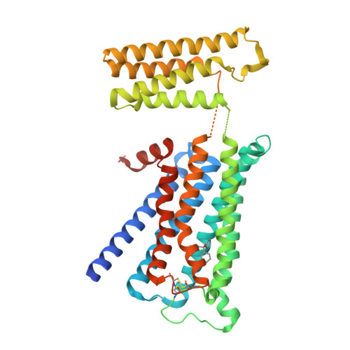
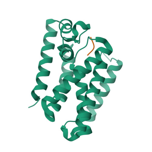
Urban, S., Cho, S. (2019) Nat Struct Mol Biol 26: 910-918 | Released | 2019-10-02 | | Method | X-RAY DIFFRACTION 2.3 Å | | Organisms | | | Macromolecule | |
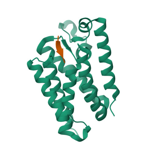
Urban, S., Cho, S. (2019) Nat Struct Mol Biol 26: 910-918 | Released | 2019-10-02 | | Method | X-RAY DIFFRACTION 2.4 Å | | Organisms | | | Macromolecule | |
|