1QXD

Structural Basis for the Potent Antisickling Effect of a Novel Class of 5-Membered Heterocyclic Aldehydic Compounds


Experimental Data Snapshot

  • Method: X-RAY DIFFRACTION
  • Resolution: 2.25 Å
  • R-Value Free: 0.274 
  • R-Value Work: 0.217 
  • R-Value Observed: 0.217 

wwPDB Validation   3D Report Full Report


Ligand Structure Quality Assessment 


This is version 1.5 of the entry. See complete history


Literature

Structural basis for the potent antisickling effect of a novel class of five-membered heterocyclic aldehydic compounds

Safo, M.K.Abdulmalik, O.Danso-Danquah, R.Burnett, J.C.Nokuri, S.Joshi, G.S.Musayev, F.N.Asakura, T.Abraham, D.J.

(2004) J Med Chem 47: 4665-4676

  • DOI: https://doi.org/10.1021/jm0498001
  • Primary Citation of Related Structures:  
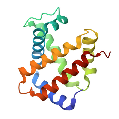
    1QXD, 1QXE

  • PubMed Abstract: 

    Naturally occurring five-membered heterocyclic aldehydes, including 5-hydroxymethyl-2-furfural, increase the oxygen affinity of hemoglobin (Hb) and strongly inhibit the sickling of homozygous sickle red blood (SS) cells. X-ray studies of Hb complexed with these compounds indicate that they form Schiff base adducts in a symmetrical fashion with the N-terminal alphaVal1 nitrogens of Hb. Interestingly, two cocrystal types were isolated during crystallization experiments with deoxygenated Hb (deoxyHb): one crystal type was composed of the low-affinity or tense (T) state Hb quaternary structure; the other crystal type was composed of high-affinity or relaxed state Hb (with a R2 quaternary structure). The R2 crystal appears to be formed as a result of the aldehydes binding to fully or partially ligated Hb in the deoxyHb solution. Repeated attempts to crystallize the compounds with liganded Hb failed, except on rare occasions when very few R state crystals were obtained. Oxygen equilibrium, high performance liquid chromatography (HPLC), antisickling, and X-ray studies suggest that the examined heterocyclic aldehydes may be acting to prevent polymerization of sickle hemoglobin (HbS) by binding to and stabilizing liganded Hb in the form of R2 and/or various relaxed state Hbs, as well as binding to and destabilizing unliganded T state Hb. The proposed mechanism may provide a general model for the antisickling effects of aldehyde containing small molecules that bind to N-terminal alphaVal1 nitrogens of Hb. The examined compounds also represent a new class of potentially therapeutic agents for treating sickle cell disease (SCD).


  • Organizational Affiliation

    Department of Medicinal Chemistry, School of Pharmacy and Institute for Structural Biology and Drug Discovery, Virginia Commonwealth University, Richmond, Virginia 23298, USA. msafo@mail2.vcu.edu


Macromolecules
Find similar proteins by:  (by identity cutoff)  |  3D Structure
Entity ID: 1
MoleculeChains Sequence LengthOrganismDetailsImage
Hemoglobin alpha chain
A, C
141Homo sapiensMutation(s): 0 
UniProt & NIH Common Fund Data Resources
Find proteins for P69905 (Homo sapiens)
Explore P69905 
Go to UniProtKB:  P69905
PHAROS:  P69905
Entity Groups  
Sequence Clusters30% Identity50% Identity70% Identity90% Identity95% Identity100% Identity
UniProt GroupP69905
Sequence Annotations
Expand
  • Reference Sequence
Find similar proteins by:  (by identity cutoff)  |  3D Structure
Entity ID: 2
MoleculeChains Sequence LengthOrganismDetailsImage
Hemoglobin beta chain
B, D
146Homo sapiensMutation(s): 0 
UniProt & NIH Common Fund Data Resources
Find proteins for P68871 (Homo sapiens)
Explore P68871 
Go to UniProtKB:  P68871
PHAROS:  P68871
GTEx:  ENSG00000244734 
Entity Groups  
Sequence Clusters30% Identity50% Identity70% Identity90% Identity95% Identity100% Identity
UniProt GroupP68871
Sequence Annotations
Expand
  • Reference Sequence
Small Molecules
Ligands 3 Unique
IDChains Name / Formula / InChI Key2D Diagram3D Interactions
HEM
Query on HEM

Download Ideal Coordinates CCD File 
F [auth A],
K [auth B],
N [auth C],
Q [auth D]
PROTOPORPHYRIN IX CONTAINING FE
C34 H32 Fe N4 O4
KABFMIBPWCXCRK-RGGAHWMASA-L
FU2
Query on FU2

Download Ideal Coordinates CCD File 
G [auth A],
O [auth C]
FURFURAL
C5 H4 O2
HYBBIBNJHNGZAN-UHFFFAOYSA-N
SO4
Query on SO4

Download Ideal Coordinates CCD File 
E [auth A]
H [auth B]
I [auth B]
J [auth B]
L [auth C]
E [auth A],
H [auth B],
I [auth B],
J [auth B],
L [auth C],
M [auth C],
P [auth D]
SULFATE ION
O4 S
QAOWNCQODCNURD-UHFFFAOYSA-L
Experimental Data & Validation

Experimental Data

  • Method: X-RAY DIFFRACTION
  • Resolution: 2.25 Å
  • R-Value Free: 0.274 
  • R-Value Work: 0.217 
  • R-Value Observed: 0.217 
  • Space Group: P 32 2 1
Unit Cell:
Length ( Å )Angle ( ˚ )
a = 91.398α = 90
b = 91.398β = 90
c = 142.003γ = 120
Software Package:
Software NamePurpose
bioteXdata collection
bioteXdata reduction
AMoREphasing
CNSrefinement
bioteXdata scaling

Structure Validation

View Full Validation Report


Ligand Structure Quality Assessment 


Entry History 

Deposition Data

Revision History  (Full details and data files)

  • Version 1.0: 2003-09-16
    Type: Initial release
  • Version 1.1: 2008-04-29
    Changes: Version format compliance
  • Version 1.2: 2011-07-13
    Changes: Version format compliance
  • Version 1.3: 2017-10-11
    Changes: Refinement description
  • Version 1.4: 2018-04-04
    Changes: Data collection
  • Version 1.5: 2023-08-23
    Changes: Data collection, Database references, Derived calculations, Refinement description