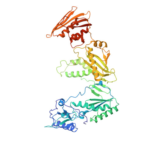
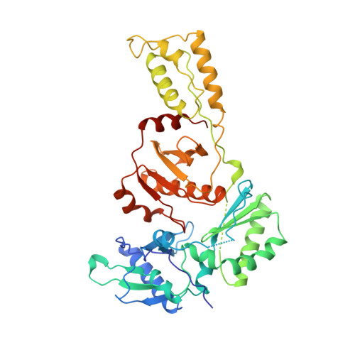
1RT5

HIV-1 REVERSE TRANSCRIPTASE COMPLEXED WITH UC10


Experimental Data Snapshot

  • Method: X-RAY DIFFRACTION
  • Resolution: 2.90 Å
  • R-Value Free: 0.291 
  • R-Value Work: 0.231 
  • R-Value Observed: 0.231 

Starting Model: experimental
View more details

wwPDB Validation   3D Report Full Report


Ligand Structure Quality Assessment 


This is version 1.3 of the entry. See complete history


Literature

Crystal structures of HIV-1 reverse transcriptase in complex with carboxanilide derivatives.

Ren, J.Esnouf, R.M.Hopkins, A.L.Warren, J.Balzarini, J.Stuart, D.I.Stammers, D.K.

(1998) Biochemistry 37: 14394-14403

  • DOI: https://doi.org/10.1021/bi981309m
  • Primary Citation of Related Structures:  
    1RT4, 1RT5, 1RT6, 1RT7

  • PubMed Abstract: 

    The carboxanilides are nonnucleoside inhibitors (NNIs) of HIV-1 reverse transcriptase (RT), of potential clinical importance. The compounds differ in potency and in their retention of potency in the face of drug resistance mutations. Whereas UC-84, the prototype compound, only weakly inhibits many RTs bearing single point resistance mutations, inhibition by UC-781 is little affected. It has been proposed that UC-38 and UC-781 may form quaternary complexes with RT at a site other than the known binding pocket of other NNIs. X-ray crystal structures of four HIV-1 RT-carboxanilide complexes (UC-10, UC-38, UC-84, and UC-781) reported here reveal that all four inhibitors bind in the usual NNI site, forming binary 1:1 complexes with RT in the absence of substrates with the amide/thioamide bond in cis conformations. For all four complexes the anilide rings of the inhibitors overlap aromatic rings of many other NNIs bound to RT. In contrast, the second rings of UC-10, UC-84, and UC-781 do not bind in equivalent positions to those of other "two-ring" NNIs such as alpha-APA or HEPT derivatives. The binding modes most closely resemble that of the structurally dissimilar NNI, Cl-TIBO, with a common hydrogen bond between each carboxanilide NH- group and the main-chain carbonyl oxygen of Lys101. The binding modes differ slightly between the UC-10/UC-781 and UC-38/UC-84 pairs of compounds, apparently related to the shorter isopropylmethanoyl substituents of the anilide rings of UC-38/UC-84, which draws these rings closer to residues Tyr181 and Tyr188. This in turn explains the differences in the effect of mutated residues on the binding of these compounds.


  • Organizational Affiliation

    Laboratory of Molecular Biophysics, Oxford, UK.


Macromolecules
Find similar proteins by:  (by identity cutoff)  |  3D Structure
Entity ID: 1
MoleculeChains Sequence LengthOrganismDetailsImage
HIV-1 REVERSE TRANSCRIPTASE560HIV-1 M:B_HXB2RMutation(s): 1 
Gene Names: POL
EC: 2.7.7.49
UniProt
Find proteins for P04585 (Human immunodeficiency virus type 1 group M subtype B (isolate HXB2))
Explore P04585 
Go to UniProtKB:  P04585
Entity Groups  
Sequence Clusters30% Identity50% Identity70% Identity90% Identity95% Identity100% Identity
UniProt GroupP04585
Sequence Annotations
Expand
  • Reference Sequence
Find similar proteins by:  (by identity cutoff)  |  3D Structure
Entity ID: 2
MoleculeChains Sequence LengthOrganismDetailsImage
HIV-1 REVERSE TRANSCRIPTASE440HIV-1 M:B_HXB2RMutation(s): 0 
Gene Names: POL
EC: 2.7.7.49
UniProt
Find proteins for P04585 (Human immunodeficiency virus type 1 group M subtype B (isolate HXB2))
Explore P04585 
Go to UniProtKB:  P04585
Entity Groups  
Sequence Clusters30% Identity50% Identity70% Identity90% Identity95% Identity100% Identity
UniProt GroupP04585
Sequence Annotations
Expand
  • Reference Sequence
Small Molecules
Ligands 2 Unique
IDChains Name / Formula / InChI Key2D Diagram3D Interactions
UC2
Query on UC2

Download Ideal Coordinates CCD File 
D [auth A]N-[4-CLORO-3-(T-BUTYLOXOME)PHENYL-2-METHYL-3-FURAN-CARBOTHIAMIDE
C17 H19 Cl N2 O2 S
PLGIIOKXCKDKEU-VXLYETTFSA-N
PO4
Query on PO4

Download Ideal Coordinates CCD File 
C [auth A]PHOSPHATE ION
O4 P
NBIIXXVUZAFLBC-UHFFFAOYSA-K
Modified Residues  1 Unique
IDChains TypeFormula2D DiagramParent
CSD
Query on CSD
A
L-PEPTIDE LINKINGC3 H7 N O4 SCYS
Experimental Data & Validation

Experimental Data

  • Method: X-RAY DIFFRACTION
  • Resolution: 2.90 Å
  • R-Value Free: 0.291 
  • R-Value Work: 0.231 
  • R-Value Observed: 0.231 
  • Space Group: P 21 21 21
Unit Cell:
Length ( Å )Angle ( ˚ )
a = 140.3α = 90
b = 111β = 90
c = 73.5γ = 90
Software Package:
Software NamePurpose
X-PLORmodel building
X-PLORrefinement
DENZOdata reduction
SCALEPACKdata scaling
X-PLORphasing

Structure Validation

View Full Validation Report


Ligand Structure Quality Assessment 


Entry History 

Deposition Data

Revision History  (Full details and data files)

  • Version 1.0: 1999-07-29
    Type: Initial release
  • Version 1.1: 2008-03-24
    Changes: Version format compliance
  • Version 1.2: 2011-07-13
    Changes: Version format compliance
  • Version 1.3: 2023-08-09
    Changes: Database references, Derived calculations, Other, Refinement description