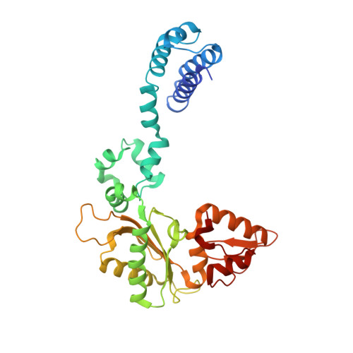
3UXO

Crystal Structure of Rat DNA Polymerase Beta Mutator I260Q Apoenzyme


Experimental Data Snapshot

  • Method: X-RAY DIFFRACTION
  • Resolution: 2.10 Å
  • R-Value Free: 0.318 
  • R-Value Work: 0.257 
  • R-Value Observed: 0.260 

wwPDB Validation   3D Report Full Report


This is version 1.2 of the entry. See complete history


Literature

Structural Changes in the Hydrophobic Hinge Region Adversely Affect the Activity and Fidelity of the I260Q Mutator DNA Polymerase beta.

Gridley, C.L.Rangarajan, S.Firbank, S.Dalal, S.Sweasy, J.B.Jaeger, J.

(2013) Biochemistry 52: 4422-4432

  • DOI: https://doi.org/10.1021/bi301368f
  • Primary Citation of Related Structures:  
    3UXN, 3UXO, 3UXP

  • PubMed Abstract: 

    The I260Q variant of DNA polymerase β is an efficient mutator polymerase with fairly indiscriminate misincorporation activities opposite all template bases. Previous modeling studies have suggested that I260Q harbors structural variations in its hinge region. Here, we present the crystal structures of wild type and I260Q rat polymerase β in the presence and absence of substrates. Both the I260Q apoenzyme structure and the closed ternary complex with double-stranded DNA and ddTTP show ordered water molecules in the hydrophobic hinge near Gln260, whereas this is not the case in the wild type polymerase. Compared to wild type polymerase β ternary complexes, there are subtle movements around residues 260, 272, 295, and 296 in the mutant. The rearrangements in this region, coupled with side chain movements in the immediate neighborhood of the dNTP-binding pocket, namely, residues 258 and 272, provide an explanation for the altered activity and fidelity profiles observed in the I260Q mutator polymerase.


  • Organizational Affiliation

    Division of Genetics, Wadsworth Center, New York State Department of Health, New Scotland Avenue, Albany, New York 12208, United States.


Macromolecules
Find similar proteins by:  (by identity cutoff)  |  3D Structure
Entity ID: 1
MoleculeChains Sequence LengthOrganismDetailsImage
DNA polymerase beta
A, B
335Rattus norvegicusMutation(s): 1 
Gene Names: Polb
EC: 2.7.7.7 (PDB Primary Data), 4.2.99 (PDB Primary Data), 4.2.99.18 (UniProt)
UniProt
Find proteins for P06766 (Rattus norvegicus)
Explore P06766 
Go to UniProtKB:  P06766
Entity Groups  
Sequence Clusters30% Identity50% Identity70% Identity90% Identity95% Identity100% Identity
UniProt GroupP06766
Sequence Annotations
Expand
  • Reference Sequence
Experimental Data & Validation

Experimental Data

  • Method: X-RAY DIFFRACTION
  • Resolution: 2.10 Å
  • R-Value Free: 0.318 
  • R-Value Work: 0.257 
  • R-Value Observed: 0.260 
  • Space Group: P 1 21 1
Unit Cell:
Length ( Å )Angle ( ˚ )
a = 77.908α = 90
b = 67.4β = 115.81
c = 82.456γ = 90
Software Package:
Software NamePurpose
CBASSdata collection
PHASERphasing
PHENIXrefinement
HKL-2000data reduction
HKL-2000data scaling

Structure Validation

View Full Validation Report


Entry History 

Deposition Data

Revision History  (Full details and data files)

  • Version 1.0: 2012-12-05
    Type: Initial release
  • Version 1.1: 2013-08-28
    Changes: Database references
  • Version 1.2: 2024-02-28
    Changes: Data collection, Database references