3VI6

Crystal Structure of human ribosomal protein L30e


Experimental Data Snapshot

  • Method: X-RAY DIFFRACTION
  • Resolution: 1.59 Å
  • R-Value Free: 0.216 
  • R-Value Work: 0.190 
  • R-Value Observed: 0.191 

wwPDB Validation   3D Report Full Report


This is version 1.3 of the entry. See complete history


Literature

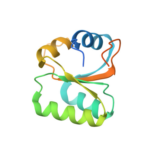
Crystallization and preliminary X-ray structure analysis of human ribosomal protein L30e

Kawaguchi, A.Ose, T.Yao, M.Tanaka, I.

(2011) Acta Crystallogr Sect F Struct Biol Cryst Commun 67: 1516-1518

  • DOI: https://doi.org/10.1107/S1744309111045131
  • Primary Citation of Related Structures:  
    3VI6

  • PubMed Abstract: 

    Many functions have been reported for the eukaryotic ribosomal protein L30e. L30e makes several inter-subunit and intra-subunit interactions with protein or RNA components of the 80S ribosome. Yeast L30e has been shown to bind to its own transcript to autoregulate expression at both the transcriptional and the translational levels. Furthermore, it has been reported that mammalian L30e is a component of the selenocysteine-incorporation machinery by binding to the selenocysteine-insertion sequence on mRNA. As high-resolution crystal structures of mammalian L30e are not available, the purification, crystallization and X-ray structure analysis of human L30e are presented here.


  • Organizational Affiliation

    Graduate School of Life Sciences, Hokkaido University, Hokkaido, Sapporo, Japan.


Macromolecules
Find similar proteins by:  (by identity cutoff)  |  3D Structure
Entity ID: 1
MoleculeChains Sequence LengthOrganismDetailsImage
60S ribosomal protein L30125Homo sapiensMutation(s): 0 
Gene Names: RPL30
UniProt & NIH Common Fund Data Resources
Find proteins for P62888 (Homo sapiens)
Explore P62888 
Go to UniProtKB:  P62888
PHAROS:  P62888
GTEx:  ENSG00000156482 
Entity Groups  
Sequence Clusters30% Identity50% Identity70% Identity90% Identity95% Identity100% Identity
UniProt GroupP62888
Sequence Annotations
Expand
  • Reference Sequence
Small Molecules
Ligands 1 Unique
IDChains Name / Formula / InChI Key2D Diagram3D Interactions
FMT
Query on FMT

Download Ideal Coordinates CCD File 
B [auth A]FORMIC ACID
C H2 O2
BDAGIHXWWSANSR-UHFFFAOYSA-N
Experimental Data & Validation

Experimental Data

  • Method: X-RAY DIFFRACTION
  • Resolution: 1.59 Å
  • R-Value Free: 0.216 
  • R-Value Work: 0.190 
  • R-Value Observed: 0.191 
  • Space Group: H 3 2
Unit Cell:
Length ( Å )Angle ( ˚ )
a = 56.282α = 90
b = 56.282β = 90
c = 203.628γ = 120
Software Package:
Software NamePurpose
SERGUIdata collection
REFMACrefinement
HKL-2000data reduction
SCALEPACKdata scaling

Structure Validation

View Full Validation Report


Entry History 

Deposition Data

Revision History  (Full details and data files)

  • Version 1.0: 2011-12-21
    Type: Initial release
  • Version 1.1: 2014-01-15
    Changes: Structure summary
  • Version 1.2: 2017-11-22
    Changes: Refinement description
  • Version 1.3: 2023-11-08
    Changes: Data collection, Database references, Derived calculations, Refinement description