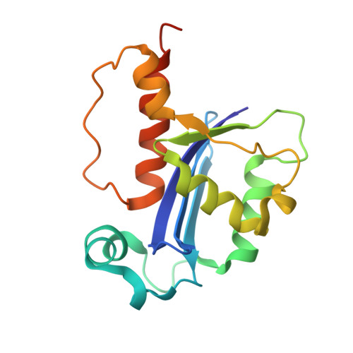
4OKE | pdb_00004oke

Structure of RNase AS, a polyadenylate-specific exoribonuclease affecting mycobacterial virulence in vivo


Experimental Data Snapshot

  • Method: X-RAY DIFFRACTION
  • Resolution: 1.70 Å
  • R-Value Free: 
    0.232 (Depositor), 0.230 (DCC) 
  • R-Value Work: 
    0.186 (Depositor), 0.190 (DCC) 
  • R-Value Observed: 
    0.188 (Depositor) 

wwPDB Validation   3D Report Full Report


Ligand Structure Quality Assessment 

Created with Raphaël 2.3.0Worse 01 BetterLigand structure goodness of fit to experimental dataBest fitted AMPClick on this verticalbar to view details

This is version 1.1 of the entry. See complete history


Literature

Structure and Function of RNase AS, a Polyadenylate-Specific Exoribonuclease Affecting Mycobacterial Virulence In Vivo

Romano, M.van de Weerd, R.Brouwer, F.C.C.Roviello, G.N.Lacroix, R.Sparrius, M.van den Brink-van Stempvoort, G.Maaskant, J.J.van der Sar, A.M.Appelmelk, B.J.Geurtsen, J.J.Berisio, R.

(2014) Structure 22: 719-730

  • DOI: https://doi.org/10.1016/j.str.2014.01.014
  • Primary Citation of Related Structures:  
    4OKE, 4OKJ, 4OKK

  • PubMed Abstract: 

    The cell-envelope of Mycobacterium tuberculosis plays a key role in bacterial virulence and antibiotic resistance. Little is known about the molecular mechanisms of regulation of cell-envelope formation. Here, we elucidate functional and structural properties of RNase AS, which modulates M. tuberculosis cell-envelope properties and strongly impacts bacterial virulence in vivo. The structure of RNase AS reveals a resemblance to RNase T from Escherichia coli, an RNase of the DEDD family involved in RNA maturation. We show that RNase AS acts as a 3'-5'-exoribonuclease that specifically hydrolyzes adenylate-containing RNA sequences. Also, crystal structures of complexes with AMP and UMP reveal the structural basis for the observed enzyme specificity. Notably, RNase AS shows a mechanism of substrate recruitment, based on the recognition of the hydrogen bond donor NH2 group of adenine. Our work opens a field for the design of drugs able to reduce bacterial virulence in vivo.


  • Organizational Affiliation

    Institute of Biostructures and Bioimaging, National Research Council, Via Mezzocannone 16, I-80134 Naples, Italy.


Macromolecules
Find similar proteins by:  (by identity cutoff)  |  3D Structure
Entity ID: 1
MoleculeChains Sequence LengthOrganismDetailsImage
3'-5' exoribonuclease Rv2179c/MT2234.1
A, B
168Mycobacterium tuberculosisMutation(s): 0 
Gene Names: Rv2179cMT2234.1
EC: 3.1.13
UniProt
Find proteins for P9WJ73 (Mycobacterium tuberculosis (strain ATCC 25618 / H37Rv))
Explore P9WJ73 
Go to UniProtKB:  P9WJ73
Entity Groups  
Sequence Clusters30% Identity50% Identity70% Identity90% Identity95% Identity100% Identity
UniProt GroupP9WJ73
Sequence Annotations
Expand
  • Reference Sequence
Experimental Data & Validation

Experimental Data

  • Method: X-RAY DIFFRACTION
  • Resolution: 1.70 Å
  • R-Value Free:  0.232 (Depositor), 0.230 (DCC) 
  • R-Value Work:  0.186 (Depositor), 0.190 (DCC) 
  • R-Value Observed: 0.188 (Depositor) 
Space Group: P 21 21 21
Unit Cell:
Length ( Å )Angle ( ˚ )
a = 42.433α = 90
b = 76.664β = 90
c = 104.372γ = 90
Software Package:
Software NamePurpose
HKL-2000data collection
SOLVEphasing
REFMACrefinement
HKL-2000data reduction
HKL-2000data scaling

Structure Validation

View Full Validation Report


Ligand Structure Quality Assessment 

Created with Raphaël 2.3.0Worse 01 BetterLigand structure goodness of fit to experimental dataBest fitted AMPClick on this verticalbar to view details

Entry History 

Deposition Data

Revision History  (Full details and data files)

  • Version 1.0: 2014-05-21
    Type: Initial release
  • Version 1.1: 2024-03-20
    Changes: Data collection, Database references, Derived calculations