4YFU

Crystal structure of open Bacillus fragment DNA polymerase bound to DNA and dTTP


Experimental Data Snapshot

  • Method: X-RAY DIFFRACTION
  • Resolution: 1.50 Å
  • R-Value Free: 0.230 
  • R-Value Work: 0.195 
  • R-Value Observed: 0.197 

wwPDB Validation   3D Report Full Report


Ligand Structure Quality Assessment 


This is version 3.0 of the entry. See complete history


Literature

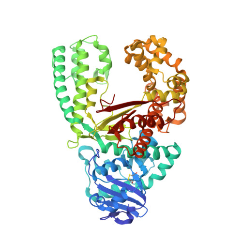
The Closing Mechanism of DNA Polymerase I at Atomic Resolution.

Miller, B.R.Beese, L.S.Parish, C.A.Wu, E.Y.

(2015) Structure 23: 1609-1620

  • DOI: https://doi.org/10.1016/j.str.2015.06.016
  • Primary Citation of Related Structures:  
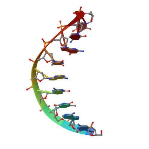
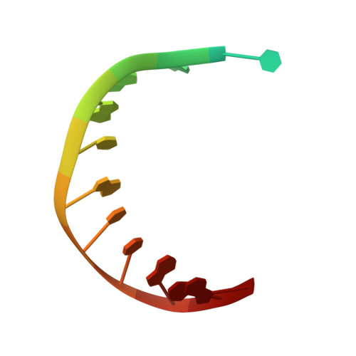
    4YFU

  • PubMed Abstract: 

    DNA polymerases must quickly and accurately distinguish between similar nucleic acids to form Watson-Crick base pairs and avoid DNA replication errors. Deoxynucleoside triphosphate (dNTP) binding to the DNA polymerase active site induces a large conformational change that is difficult to characterize experimentally on an atomic level. Here, we report an X-ray crystal structure of DNA polymerase I bound to DNA in the open conformation with a dNTP present in the active site. We use this structure to computationally simulate the open to closed transition of DNA polymerase in the presence of a Watson-Crick base pair. Our microsecond simulations allowed us to characterize the key steps involved in active site assembly, and propose the sequence of events involved in the prechemistry steps of DNA polymerase catalysis. They also reveal new features of the polymerase mechanism, such as a conserved histidine as a potential proton acceptor from the primer 3'-hydroxyl.


  • Organizational Affiliation

    Department of Biology, University of Richmond, 28 Westhampton Way, Richmond, VA 23173, USA; Department of Chemistry, University of Richmond, 28 Westhampton Way, Richmond, VA 23173, USA.


Macromolecules

Find similar proteins by:  (by identity cutoff)  |  3D Structure
Entity ID: 1
MoleculeChains Sequence LengthOrganismDetailsImage
DNA polymeraseA,
B [auth D]
580Geobacillus stearothermophilusMutation(s): 2 
EC: 2.7.7.7
UniProt
Find proteins for E1C9K5 (Geobacillus stearothermophilus)
Explore E1C9K5 
Go to UniProtKB:  E1C9K5
Entity Groups  
Sequence Clusters30% Identity50% Identity70% Identity90% Identity95% Identity100% Identity
UniProt GroupE1C9K5
Sequence Annotations
Expand
  • Reference Sequence

Find similar nucleic acids by:  Sequence   |   3D Structure  

Entity ID: 2
MoleculeChains LengthOrganismImage
Primer DNAC [auth B],
E
9synthetic construct
Sequence Annotations
Expand
  • Reference Sequence

Find similar nucleic acids by:  Sequence   |   3D Structure  

Entity ID: 3
MoleculeChains LengthOrganismImage
Template DNAD [auth C],
F
14synthetic construct
Sequence Annotations
Expand
  • Reference Sequence
Oligosaccharides

Help

Entity ID: 4
MoleculeChains Length2D Diagram Glycosylation3D Interactions
beta-D-fructofuranose-(2-1)-alpha-D-glucopyranose
G, H
2N/A
Glycosylation Resources
GlyTouCan:  G05551OP
GlyCosmos:  G05551OP
Small Molecules
Ligands 2 Unique
IDChains Name / Formula / InChI Key2D Diagram3D Interactions
TTP
Query on TTP

Download Ideal Coordinates CCD File 
I [auth A]THYMIDINE-5'-TRIPHOSPHATE
C10 H17 N2 O14 P3
NHVNXKFIZYSCEB-XLPZGREQSA-N
SO4
Query on SO4

Download Ideal Coordinates CCD File 
J [auth D]SULFATE ION
O4 S
QAOWNCQODCNURD-UHFFFAOYSA-L
Modified Residues  1 Unique
IDChains TypeFormula2D DiagramParent
CME
Query on CME
A,
B [auth D]
L-PEPTIDE LINKINGC5 H11 N O3 S2CYS
Biologically Interesting Molecules (External Reference) 1 Unique
Experimental Data & Validation

Experimental Data

  • Method: X-RAY DIFFRACTION
  • Resolution: 1.50 Å
  • R-Value Free: 0.230 
  • R-Value Work: 0.195 
  • R-Value Observed: 0.197 
  • Space Group: P 21 21 21
Unit Cell:
Length ( Å )Angle ( ˚ )
a = 93.23α = 90
b = 108.1β = 90
c = 151.34γ = 90
Software Package:
Software NamePurpose
REFMACrefinement
DENZOdata reduction
XSCALEdata scaling
PDB_EXTRACTdata extraction
REFMACphasing
Cootmodel building

Structure Validation

View Full Validation Report


Ligand Structure Quality Assessment 


Entry History & Funding Information

Deposition Data

  • Released Date: 2015-07-22 
  • Deposition Author(s): Wu, E.Y.

Funding OrganizationLocationGrant Number
Jeffress TrustUnited States--

Revision History  (Full details and data files)

  • Version 1.0: 2015-07-22
    Type: Initial release
  • Version 1.1: 2015-08-12
    Changes: Refinement description
  • Version 1.2: 2015-09-16
    Changes: Database references
  • Version 2.0: 2020-07-29
    Type: Remediation
    Reason: Carbohydrate remediation
    Changes: Atomic model, Data collection, Database references, Derived calculations, Non-polymer description, Structure summary
  • Version 3.0: 2022-04-13
    Changes: Advisory, Atomic model, Data collection, Database references, Derived calculations, Structure summary