4Z4W | pdb_00004z4w

Crystal structure of GII.10 P domain in complex with 4.7mM fucose


Experimental Data Snapshot

  • Method: X-RAY DIFFRACTION
  • Resolution: 1.80 Å
  • R-Value Free: 
    0.191 (Depositor), 0.190 (DCC) 
  • R-Value Work: 
    0.155 (Depositor), 0.160 (DCC) 
  • R-Value Observed: 
    0.156 (Depositor) 

Starting Model: experimental
View more details

wwPDB Validation   3D Report Full Report


Ligand Structure Quality Assessment 

Created with Raphaël 2.3.0Worse 01 BetterLigand structure goodness of fit to experimental dataBest fitted FULClick on this verticalbar to view details

This is version 1.3 of the entry. See complete history


Literature

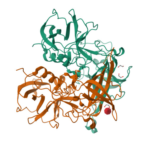
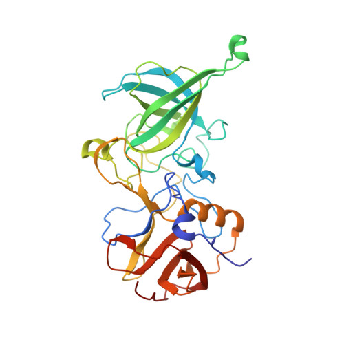
The sweet quartet: Binding of fucose to the norovirus capsid.

Koromyslova, A.D.Leuthold, M.M.Bowler, M.W.Hansman, G.S.

(2015) Virology 483: 203-208

  • DOI: https://doi.org/10.1016/j.virol.2015.04.006
  • Primary Citation of Related Structures:  
    4Z4R, 4Z4S, 4Z4T, 4Z4U, 4Z4V, 4Z4W, 4Z4Y, 4Z4Z

  • PubMed Abstract: 

    Human noroviruses bind histo-blood group antigens (HBGAs) and this interaction is thought to be important for an infection. We identified two additional fucose-binding pockets (termed fucose-3/4 sites) on a genogroup II human (GII.10) norovirus-protruding (P) dimer using X-ray crystallography. Fucose-3/4 sites were located between two previously determined HBGA binding pockets (termed fucose-1/2 sites). We found that four fucose molecules were capable of binding altogether at fucose-1/2/3/4 sites on the P dimer, though the fucose molecules bound in a dose-dependent and step-wise manner. We also showed that HBGA B-trisaccharide molecules bound in a similar way at the fucose-1/2 sites. Interestingly, we discovered that the monomers of the P dimer were asymmetrical in an unliganded state and when a single B-trisaccharide molecule bound, but were symmetrical when two B-trisaccharide molecules bound. We postulate that the symmetrical dimers might favor HBGA binding interactions at fucose-1/2 sites.


  • Organizational Affiliation

    Schaller Research Group at the University of Heidelberg and the DKFZ, Heidelberg 69120, Germany; Department of Infectious Diseases, Virology, University of Heidelberg, Heidelberg 69120, Germany.


Macromolecules
Find similar proteins by:  (by identity cutoff)  |  3D Structure
Entity ID: 1
MoleculeChains Sequence LengthOrganismDetailsImage
Capsid protein
A, B
315Norovirus GII.10Mutation(s): 0 
UniProt
Find proteins for Q5F4T5 (Norwalk virus)
Explore Q5F4T5 
Go to UniProtKB:  Q5F4T5
Entity Groups  
Sequence Clusters30% Identity50% Identity70% Identity90% Identity95% Identity100% Identity
UniProt GroupQ5F4T5
Sequence Annotations
Expand
  • Reference Sequence
Experimental Data & Validation

Experimental Data

  • Method: X-RAY DIFFRACTION
  • Resolution: 1.80 Å
  • R-Value Free:  0.191 (Depositor), 0.190 (DCC) 
  • R-Value Work:  0.155 (Depositor), 0.160 (DCC) 
  • R-Value Observed: 0.156 (Depositor) 
Space Group: P 1 21 1
Unit Cell:
Length ( Å )Angle ( ˚ )
a = 65.29α = 90
b = 79.344β = 100.78
c = 69.576γ = 90
Software Package:
Software NamePurpose
PHASERphasing
PHENIXrefinement
PDB_EXTRACTdata extraction
XDSdata reduction

Structure Validation

View Full Validation Report


Ligand Structure Quality Assessment 

Created with Raphaël 2.3.0Worse 01 BetterLigand structure goodness of fit to experimental dataBest fitted FULClick on this verticalbar to view details

Entry History 

Deposition Data

Revision History  (Full details and data files)

  • Version 1.0: 2015-05-27
    Type: Initial release
  • Version 1.1: 2015-06-03
    Changes: Database references
  • Version 1.2: 2020-07-29
    Type: Remediation
    Reason: Carbohydrate remediation
    Changes: Data collection, Derived calculations, Structure summary
  • Version 1.3: 2024-01-10
    Changes: Data collection, Database references, Refinement description, Structure summary