


Crystal structure of an intact type IV self-sufficient cytochrome P450 monooxygenase
Gong, R., Wu, L.J., Zhang, Y., Liu, Z., Dou, S., Zhang, R.G., Xu, J.H., Tang, C., Zhou, J.H.To be published.
Experimental Data Snapshot
Entity ID: 1 | |||||
|---|---|---|---|---|---|
| Molecule | Chains | Sequence Length | Organism | Details | Image |
| Cytochrome P450 monooxygenase | 786 | Rhodococcus sp. ECU0066 | Mutation(s): 0 | 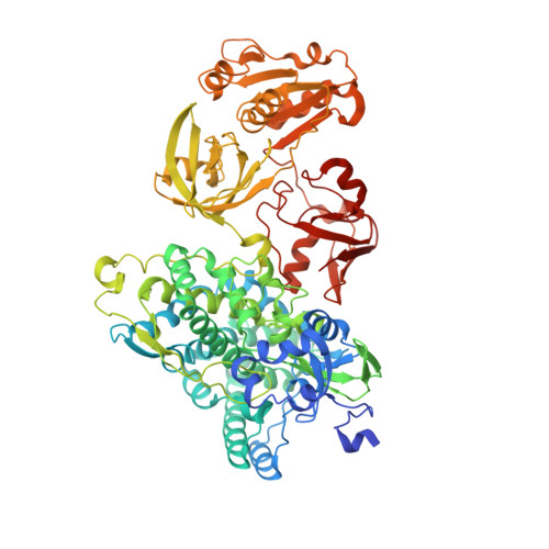 | |
UniProt | |||||
Find proteins for C7SFP5 (Rhodococcus sp. ECU0066) Explore C7SFP5 Go to UniProtKB: C7SFP5 | |||||
Entity Groups | |||||
| Sequence Clusters | 30% Identity50% Identity70% Identity90% Identity95% Identity100% Identity | ||||
| UniProt Group | C7SFP5 | ||||
Sequence AnnotationsExpand | |||||
| |||||
| Ligands 3 Unique | |||||
|---|---|---|---|---|---|
| ID | Chains | Name / Formula / InChI Key | 2D Diagram | 3D Interactions | |
| HEM (Subject of Investigation/LOI) Query on HEM | C [auth A] | PROTOPORPHYRIN IX CONTAINING FE C34 H32 Fe N4 O4 KABFMIBPWCXCRK-RGGAHWMASA-L |  | ||
| FMN Query on FMN | B [auth A] | FLAVIN MONONUCLEOTIDE C17 H21 N4 O9 P FVTCRASFADXXNN-SCRDCRAPSA-N |  | ||
| FES (Subject of Investigation/LOI) Query on FES | D [auth A] | FE2/S2 (INORGANIC) CLUSTER Fe2 S2 NIXDOXVAJZFRNF-UHFFFAOYSA-N |  | ||
| Length ( Å ) | Angle ( ˚ ) |
|---|---|
| a = 131.762 | α = 90 |
| b = 55.484 | β = 98.12 |
| c = 135.96 | γ = 90 |
| Software Name | Purpose |
|---|---|
| PHENIX | refinement |
| XSCALE | data scaling |
| PDB_EXTRACT | data extraction |
| XDS | data reduction |
| PHASER | phasing |


