


Mycobacterial sensor histidine kinase MprB
Li, J., Korotkova, N., Korotkov, K.V.To be published.
Experimental Data Snapshot
Starting Model: experimental
View more details
Entity ID: 1 | |||||
|---|---|---|---|---|---|
| Molecule | Chains | Sequence Length | Organism | Details | Image |
| Signal transduction histidine-protein kinase/phosphatase mprB | A [auth C], B [auth D], C [auth A], D [auth B] | 158 | Mycolicibacterium hassiacum DSM 44199 | Mutation(s): 0 Gene Names: mprB, C731_3480 EC: 3.1.3 (PDB Primary Data), 2.7.13.3 (UniProt) | 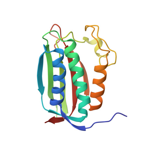 |
UniProt | |||||
Find proteins for K5BDW2 (Mycolicibacterium hassiacum (strain DSM 44199 / CIP 105218 / JCM 12690 / 3849)) Explore K5BDW2 Go to UniProtKB: K5BDW2 | |||||
Entity Groups | |||||
| Sequence Clusters | 30% Identity50% Identity70% Identity90% Identity95% Identity100% Identity | ||||
| UniProt Group | K5BDW2 | ||||
Sequence AnnotationsExpand | |||||
| |||||
| Ligands 2 Unique | |||||
|---|---|---|---|---|---|
| ID | Chains | Name / Formula / InChI Key | 2D Diagram | 3D Interactions | |
| ATP (Subject of Investigation/LOI) Query on ATP | E [auth C], G [auth D], I [auth A], K [auth B] | ADENOSINE-5'-TRIPHOSPHATE C10 H16 N5 O13 P3 ZKHQWZAMYRWXGA-KQYNXXCUSA-N |  | ||
| MG (Subject of Investigation/LOI) Query on MG | F [auth C], H [auth D], J [auth A], L [auth B] | MAGNESIUM ION Mg JLVVSXFLKOJNIY-UHFFFAOYSA-N |  | ||
| Length ( Å ) | Angle ( ˚ ) |
|---|---|
| a = 40.41 | α = 102.78 |
| b = 61.63 | β = 104.47 |
| c = 67.22 | γ = 109.08 |
| Software Name | Purpose |
|---|---|
| XSCALE | data scaling |
| PHENIX | refinement |
| PDB_EXTRACT | data extraction |
| XDS | data reduction |
| BALBES | phasing |
| Funding Organization | Location | Grant Number |
|---|---|---|
| National Institutes of Health/National Institute Of Allergy and Infectious Diseases (NIH/NIAID) | United States | R01AI119022 |
| National Institutes of Health/National Institute of General Medical Sciences (NIH/NIGMS) | United States | P30GM110787 |


