Cell-free protein crystallization for nanocrystal structure determination.
Abe, S., Tanaka, J., Kojima, M., Kanamaru, S., Hirata, K., Yamashita, K., Kobayashi, A., Ueno, T.(2022) Sci Rep 12: 16031-16031
- PubMed: 36192567
- DOI: https://doi.org/10.1038/s41598-022-19681-9
- Primary Citation of Related Structures:
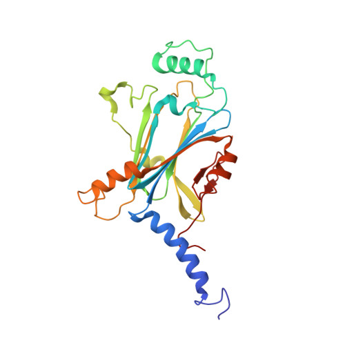
7XHR, 7XHS, 7XWS - PubMed Abstract:
In-cell protein crystallization (ICPC) has been investigated as a technique to support the advancement of structural biology because it does not require protein purification and a complicated crystallization process. However, only a few protein structures have been reported because these crystals formed incidentally in living cells and are insufficient in size and quality for structure analysis. Here, we have developed a cell-free protein crystallization (CFPC) method, which involves direct protein crystallization using cell-free protein synthesis. We have succeeded in crystallization and structure determination of nano-sized polyhedra crystal (PhC) at a high resolution of 1.80 Å. Furthermore, nanocrystals were synthesized at a reaction scale of only 20 μL using the dialysis method, enabling structural analysis at a resolution of 1.95 Å. To further demonstrate the potential of CFPC, we attempted to determine the structure of crystalline inclusion protein A (CipA), whose structure had not yet been determined. We added chemical reagents as a twinning inhibitor to the CFPC solution, which enabled us to determine the structure of CipA at 2.11 Å resolution. This technology greatly expands the high-throughput structure determination method of unstable, low-yield, fusion, and substrate-biding proteins that have been difficult to analyze with conventional methods.
- School of Life Science and Technology, Tokyo Institute of Technology, Nagatsuta-cho 4259, Midori-ku, Yokohama, 226-8501, Japan. saabe@bio.titech.ac.jp.
Organizational Affiliation: