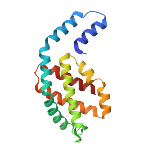
8OR7 | pdb_00008or7

Structure of a far-red induced allophycocyanin from Chroococcidiopsis thermalis sp. PCC 7203


Experimental Data Snapshot

  • Method: X-RAY DIFFRACTION
  • Resolution: 2.80 Å
  • R-Value Free: 
    0.240 (Depositor), 0.240 (DCC) 
  • R-Value Work: 
    0.178 (Depositor), 0.180 (DCC) 
  • R-Value Observed: 
    0.181 (Depositor) 

Starting Models: experimental
View more details

wwPDB Validation   3D Report Full Report


Ligand Structure Quality Assessment 


This is version 1.2 of the entry. See complete history


Literature

Crystallographic and biochemical analyses of a far-red allophycocyanin to address the mechanism of the super-red-shift.

Zhou, L.J.Hoppner, A.Wang, Y.Q.Hou, J.Y.Scheer, H.Zhao, K.H.

(2024) Photosynth Res 162: 171-185

  • DOI: https://doi.org/10.1007/s11120-023-01066-2
  • Primary Citation of Related Structures:  
    8OR7

  • PubMed Abstract: 

    Far-red absorbing allophycocyanins (APC), identified in cyanobacteria capable of FRL photoacclimation (FaRLiP) and low-light photoacclimation (LoLiP), absorb far-red light, functioning in energy transfer as light-harvesting proteins. We report an optimized method to obtain high purity far-red absorbing allophycocyanin B, AP-B2, of Chroococcidiopsis thermalis sp. PCC7203 by synthesis in Escherichia coli and an improved purification protocol. The crystal structure of the trimer, (PCB-ApcD5/PCB-ApcB2) 3 , has been resolved to 2.8 Å. The main difference to conventional APCs absorbing in the 650-670 nm range is a largely flat chromophore with the co-planarity extending, in particular, from rings BCD to ring A. This effectively extends the conjugation system of PCB and contributes to the super-red-shifted absorption of the α-subunit (λ max  = 697 nm). On complexation with the β-subunit, it is even further red-shifted (λ max, absorption  = 707 nm, λ max, emission  = 721 nm). The relevance of ring A for this shift is supported by mutagenesis data. A variant of the α-subunit, I123M, has been generated that shows an intense FR-band already in the absence of the β-subunit, a possible model is discussed. Two additional mechanisms are known to red-shift the chromophore spectrum: lactam-lactim tautomerism and deprotonation of the chromophore that both mechanisms appear inconsistent with our data, leaving this question unresolved.


  • Organizational Affiliation
    • National Key Laboratory of Agricultural Microbiology, Huazhong Agricultural University, Wuhan, 430070, The People's Republic of China.

Macromolecules
Find similar proteins by:  (by identity cutoff)  |  3D Structure
Entity ID: 1
MoleculeChains Sequence LengthOrganismDetailsImage
Allophycocyanin beta subunit apoproteinA [auth B],
B [auth D]
169Chroococcidiopsis thermalisMutation(s): 0 
Gene Names: Chro_1033
UniProt
Find proteins for K9TVG8 (Chroococcidiopsis thermalis (strain PCC 7203))
Explore K9TVG8 
Go to UniProtKB:  K9TVG8
Entity Groups  
Sequence Clusters30% Identity50% Identity70% Identity90% Identity95% Identity100% Identity
UniProt GroupK9TVG8
Sequence Annotations
Expand
  • Reference Sequence
Find similar proteins by:  (by identity cutoff)  |  3D Structure
Entity ID: 2
MoleculeChains Sequence LengthOrganismDetailsImage
PhycocyaninC [auth A],
D [auth C]
159Chroococcidiopsis thermalisMutation(s): 0 
Gene Names: Chro_1032
UniProt
Find proteins for K9TX42 (Chroococcidiopsis thermalis (strain PCC 7203))
Explore K9TX42 
Go to UniProtKB:  K9TX42
Entity Groups  
Sequence Clusters30% Identity50% Identity70% Identity90% Identity95% Identity100% Identity
UniProt GroupK9TX42
Sequence Annotations
Expand
  • Reference Sequence
Small Molecules
Ligands 3 Unique
IDChains Name / Formula / InChI Key2D Diagram3D Interactions
CYC (Subject of Investigation/LOI)
Query on CYC

Download Ideal Coordinates CCD File 
E [auth B],
H [auth D],
I [auth A],
L [auth C]
PHYCOCYANOBILIN
C33 H40 N4 O6
VXTXPYZGDQPMHK-GMXXPEQVSA-N
SO4
Query on SO4

Download Ideal Coordinates CCD File 
G [auth B],
M [auth C],
O [auth C]
SULFATE ION
O4 S
QAOWNCQODCNURD-UHFFFAOYSA-L
K
Query on K

Download Ideal Coordinates CCD File 
F [auth B],
J [auth A],
K [auth A],
N [auth C]
POTASSIUM ION
K
NPYPAHLBTDXSSS-UHFFFAOYSA-N
Experimental Data & Validation

Experimental Data

  • Method: X-RAY DIFFRACTION
  • Resolution: 2.80 Å
  • R-Value Free:  0.240 (Depositor), 0.240 (DCC) 
  • R-Value Work:  0.178 (Depositor), 0.180 (DCC) 
  • R-Value Observed: 0.181 (Depositor) 
Space Group: P 63
Unit Cell:
Length ( Å )Angle ( ˚ )
a = 130.54α = 90
b = 130.54β = 90
c = 106.496γ = 120
Software Package:
Software NamePurpose
REFMACrefinement
xia2data reduction
pointlessdata scaling
ARP/wARPphasing

Structure Validation

View Full Validation Report


Ligand Structure Quality Assessment 


Entry History & Funding Information

Deposition Data


Funding OrganizationLocationGrant Number
National Natural Science Foundation of China (NSFC)China31861143029

Revision History  (Full details and data files)

  • Version 1.0: 2024-03-20
    Type: Initial release
  • Version 1.1: 2024-11-06
    Changes: Structure summary
  • Version 1.2: 2024-12-11
    Changes: Database references