8F26

Structure of a 60mer DegP cage bound to the client protein hTRF1


Experimental Data Snapshot

  • Method: ELECTRON MICROSCOPY
  • Resolution: 9.70 Å
  • Aggregation State: PARTICLE 
  • Reconstruction Method: SINGLE PARTICLE 

wwPDB Validation   3D Report Full Report


This is version 1.2 of the entry. See complete history


Literature

Flexible Client-Dependent Cages in the Assembly Landscape of the Periplasmic Protease-Chaperone DegP.

Harkness, R.W.Ripstein, Z.A.Di Trani, J.M.Kay, L.E.

(2023) J Am Chem Soc 145: 13015-13026

  • DOI: https://doi.org/10.1021/jacs.2c11849
  • Primary Citation of Related Structures:  
    8F0A, 8F0U, 8F1T, 8F1U, 8F21, 8F26

  • PubMed Abstract: 

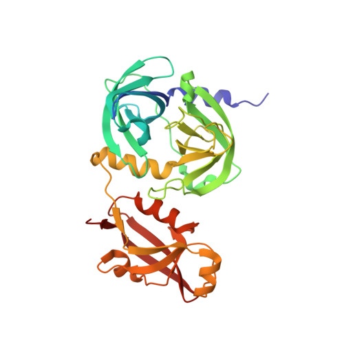
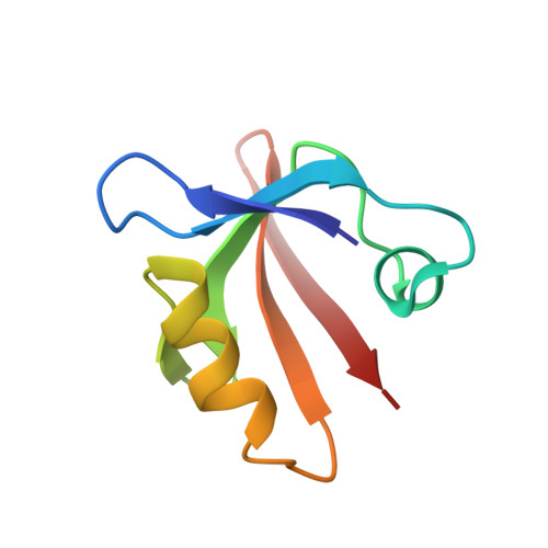
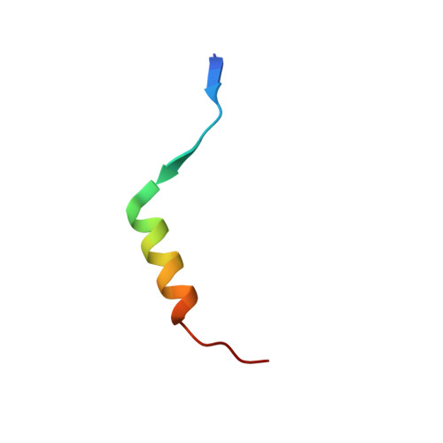
    The periplasmic protein DegP, which is implicated in virulence factor transport leading to pathogenicity, is a bi-functional protease and chaperone that helps to maintain protein homeostasis in Gram-negative bacteria and is essential to bacterial survival under stress conditions. To perform these functions, DegP captures clients inside cage-like structures, which we have recently shown to form through the reorganization of high-order preformed apo oligomers, consisting of trimeric building blocks, that are structurally distinct from client-bound cages. Our previous studies suggested that these apo oligomers may allow DegP to encapsulate clients of various sizes under protein folding stresses by forming ensembles that can include extremely large cage particles, but how this occurs remains an open question. To explore the relation between cage and substrate sizes, we engineered a series of DegP clients of increasing hydrodynamic radii and analyzed their influence on DegP cage formation. We used dynamic light scattering and cryogenic electron microscopy to characterize the hydrodynamic properties and structures of the DegP cages that are adopted in response to each client. We present a series of density maps and structural models that include those for novel particles of approximately 30 and 60 monomers. Key interactions between DegP trimers and the bound clients that stabilize the cage assemblies and prime the clients for catalysis are revealed. We also provide evidence that DegP can form cages which approach subcellular organelles in terms of size.


  • Organizational Affiliation

    Department of Biochemistry, University of Toronto, Toronto M5S 1A8, Canada.


Macromolecules
Find similar proteins by:  (by identity cutoff)  |  3D Structure
Entity ID: 1
MoleculeChains Sequence LengthOrganismDetailsImage
Periplasmic serine endoprotease DegP348Escherichia coli K-12Mutation(s): 0 
Gene Names: degPhtrAptdb0161JW0157
EC: 3.4.21.107
UniProt
Find proteins for P0C0V0 (Escherichia coli (strain K12))
Explore P0C0V0 
Go to UniProtKB:  P0C0V0
Entity Groups  
Sequence Clusters30% Identity50% Identity70% Identity90% Identity95% Identity100% Identity
UniProt GroupP0C0V0
Sequence Annotations
Expand
  • Reference Sequence
Find similar proteins by:  (by identity cutoff)  |  3D Structure
Entity ID: 2
MoleculeChains Sequence LengthOrganismDetailsImage
Periplasmic serine endoprotease DegPB [auth D]75Escherichia coli K-12Mutation(s): 0 
Gene Names: degPhtrAptdb0161JW0157
EC: 3.4.21.107
UniProt
Find proteins for P0C0V0 (Escherichia coli (strain K12))
Explore P0C0V0 
Go to UniProtKB:  P0C0V0
Entity Groups  
Sequence Clusters30% Identity50% Identity70% Identity90% Identity95% Identity100% Identity
UniProt GroupP0C0V0
Sequence Annotations
Expand
  • Reference Sequence
Find similar proteins by:  (by identity cutoff)  |  3D Structure
Entity ID: 3
MoleculeChains Sequence LengthOrganismDetailsImage
Telomeric repeat-binding factor 1C [auth a]27Homo sapiensMutation(s): 0 
Gene Names: TERF1PIN2TRBF1TRFTRF1
UniProt & NIH Common Fund Data Resources
Find proteins for P54274 (Homo sapiens)
Explore P54274 
Go to UniProtKB:  P54274
PHAROS:  P54274
GTEx:  ENSG00000147601 
Entity Groups  
Sequence Clusters30% Identity50% Identity70% Identity90% Identity95% Identity100% Identity
UniProt GroupP54274
Sequence Annotations
Expand
  • Reference Sequence
Experimental Data & Validation

Experimental Data

  • Method: ELECTRON MICROSCOPY
  • Resolution: 9.70 Å
  • Aggregation State: PARTICLE 
  • Reconstruction Method: SINGLE PARTICLE 

Structure Validation

View Full Validation Report


Entry History & Funding Information

Deposition Data


Funding OrganizationLocationGrant Number
Canadian Institutes of Health Research (CIHR)CanadaFDN-503573

Revision History  (Full details and data files)

  • Version 1.0: 2022-11-23
    Type: Initial release
  • Version 1.1: 2023-06-21
    Changes: Database references
  • Version 1.2: 2023-07-05
    Changes: Database references