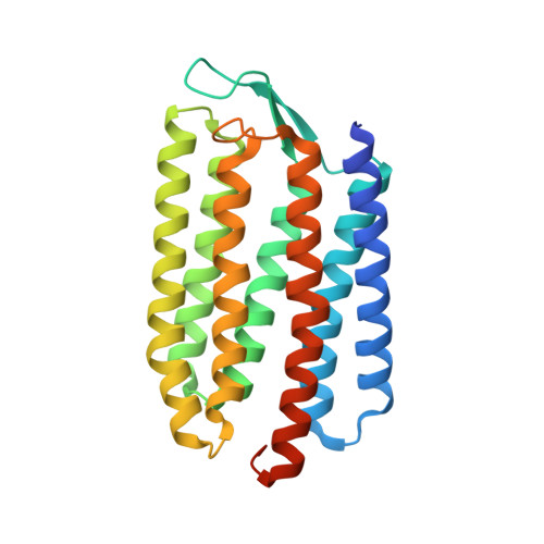
8H79 | pdb_00008h79

The crystal structure of cyanorhodopsin-II (CyR-II) P7104R from Nodosilinea nodulosa PCC 7104


Experimental Data Snapshot

  • Method: X-RAY DIFFRACTION
  • Resolution: 2.07 Å
  • R-Value Free: 
    0.248 (Depositor), 0.250 (DCC) 
  • R-Value Work: 
    0.203 (Depositor), 0.200 (DCC) 
  • R-Value Observed: 
    0.207 (Depositor) 

wwPDB Validation   3D Report Full Report


Ligand Structure Quality Assessment 


This is version 1.1 of the entry. See complete history


Literature

Cyanorhodopsin-II represents a yellow-absorbing proton-pumping rhodopsin clade within cyanobacteria.

Hasegawa-Takano, M.Hosaka, T.Kojima, K.Nishimura, Y.Kurihara, M.Nakajima, Y.Ishizuka-Katsura, Y.Kimura-Someya, T.Shirouzu, M.Sudo, Y.Yoshizawa, S.

(2024) ISME J 18

  • DOI: https://doi.org/10.1093/ismejo/wrae175
  • Primary Citation of Related Structures:  
    8H79

  • PubMed Abstract: 

    Microbial rhodopsins are prevalent in many cyanobacterial groups as a light-energy-harvesting system in addition to the photosynthetic system. It has been suggested that this dual system allows efficient capture of sunlight energy using complementary ranges of absorption wavelengths. However, the diversity of cyanobacterial rhodopsins, particularly in accumulated metagenomic data, remains underexplored. Here, we used a metagenomic mining approach, which led to the identification of a novel rhodopsin clade unique to cyanobacteria, cyanorhodopsin-II (CyR-II). CyR-IIs function as light-driven outward H+ pumps. CyR-IIs, together with previously identified cyanorhodopsins (CyRs) and cyanobacterial halorhodopsins (CyHRs), constitute cyanobacterial ion-pumping rhodopsins (CyipRs), a phylogenetically distinct family of rhodopsins. The CyR-II clade is further divided into two subclades, YCyR-II and GCyR-II, based on their specific absorption wavelength. YCyR-II absorbed yellow light (λmax = 570 nm), whereas GCyR-II absorbed green light (λmax = 550 nm). X-ray crystallography and mutational analysis revealed that the difference in absorption wavelengths is attributable to slight changes in the side chain structure near the retinal chromophore. The evolutionary trajectory of cyanobacterial rhodopsins suggests that the function and light-absorbing range of these rhodopsins have been adapted to a wide range of habitats with variable light and environmental conditions. Collectively, these findings shed light on the importance of rhodopsins in the evolution and environmental adaptation of cyanobacteria.


  • Organizational Affiliation
    • Atmosphere and Ocean Research Institute, The University of Tokyo, Chiba 277-8564, Japan.

Macromolecules
Find similar proteins by:  (by identity cutoff)  |  3D Structure
Entity ID: 1
MoleculeChains Sequence LengthOrganismDetailsImage
cyanorhodopsin-II (CyR-II) P7104R256Nodosilinea nodulosa PCC 7104Mutation(s): 0 
Membrane Entity: Yes 
UniProt
Find proteins for A0AAJ6N6B2 (Nodosilinea nodulosa PCC 7104)
Explore A0AAJ6N6B2 
Go to UniProtKB:  A0AAJ6N6B2
Entity Groups  
Sequence Clusters30% Identity50% Identity70% Identity90% Identity95% Identity100% Identity
UniProt GroupA0AAJ6N6B2
Sequence Annotations
Expand
  • Reference Sequence
Small Molecules
Ligands 8 Unique
IDChains Name / Formula / InChI Key2D Diagram3D Interactions
RET
Query on RET

Download Ideal Coordinates CCD File 
B [auth A]RETINAL
C20 H28 O
NCYCYZXNIZJOKI-OVSJKPMPSA-N
R16
Query on R16

Download Ideal Coordinates CCD File 
L [auth A]HEXADECANE
C16 H34
DCAYPVUWAIABOU-UHFFFAOYSA-N
C14
Query on C14

Download Ideal Coordinates CCD File 
J [auth A],
K [auth A]
TETRADECANE
C14 H30
BGHCVCJVXZWKCC-UHFFFAOYSA-N
PG4
Query on PG4

Download Ideal Coordinates CCD File 
C [auth A]TETRAETHYLENE GLYCOL
C8 H18 O5
UWHCKJMYHZGTIT-UHFFFAOYSA-N
OCT
Query on OCT

Download Ideal Coordinates CCD File 
G [auth A],
H [auth A],
I [auth A]
N-OCTANE
C8 H18
TVMXDCGIABBOFY-UHFFFAOYSA-N
SO4
Query on SO4

Download Ideal Coordinates CCD File 
M [auth A]SULFATE ION
O4 S
QAOWNCQODCNURD-UHFFFAOYSA-L
HEX
Query on HEX

Download Ideal Coordinates CCD File 
D [auth A],
E [auth A],
F [auth A]
HEXANE
C6 H14
VLKZOEOYAKHREP-UHFFFAOYSA-N
CL
Query on CL

Download Ideal Coordinates CCD File 
N [auth A]CHLORIDE ION
Cl
VEXZGXHMUGYJMC-UHFFFAOYSA-M
Experimental Data & Validation

Experimental Data

  • Method: X-RAY DIFFRACTION
  • Resolution: 2.07 Å
  • R-Value Free:  0.248 (Depositor), 0.250 (DCC) 
  • R-Value Work:  0.203 (Depositor), 0.200 (DCC) 
  • R-Value Observed: 0.207 (Depositor) 
Space Group: P 3 2 1
Unit Cell:
Length ( Å )Angle ( ˚ )
a = 63.15α = 90
b = 63.15β = 90
c = 117.76γ = 120
Software Package:
Software NamePurpose
PHENIXrefinement
XSCALEdata scaling
PDB_EXTRACTdata extraction
XDSdata reduction
PHASERphasing

Structure Validation

View Full Validation Report


Ligand Structure Quality Assessment 


Entry History & Funding Information

Deposition Data


Funding OrganizationLocationGrant Number
Japan Society for the Promotion of Science (JSPS)Japan17K07324
Japan Society for the Promotion of Science (JSPS)Japan20H05450

Revision History  (Full details and data files)

  • Version 1.0: 2023-10-25
    Type: Initial release
  • Version 1.1: 2024-11-13
    Changes: Database references, Structure summary