8I8E

Acyl-ACP synthetase structure bound to C18:1-ACP


Experimental Data Snapshot

  • Method: ELECTRON MICROSCOPY
  • Resolution: 2.63 Å
  • Aggregation State: PARTICLE 
  • Reconstruction Method: SINGLE PARTICLE 

wwPDB Validation   3D Report Full Report


This is version 2.1 of the entry. See complete history


Literature

Structure and catalytic mechanism of exogenous fatty acid recycling by AasS, a versatile acyl-ACP synthetase.

Huang, H.Wang, C.Chang, S.Cui, T.Xu, Y.Huang, M.Zhang, H.Zhou, C.Zhang, X.Feng, Y.

(2025) Nat Struct Mol Biol 

  • DOI: https://doi.org/10.1038/s41594-024-01464-7
  • Primary Citation of Related Structures:  
    8I35, 8I3I, 8I49, 8I51, 8I6M, 8I8D, 8I8E

  • PubMed Abstract: 

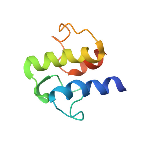
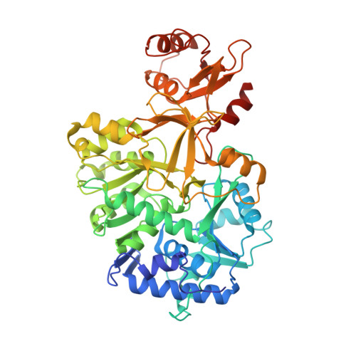
    Fatty acids (FAs) are essential building blocks for all the domains of life, of which bacterial de novo synthesis, called type II FA synthesis (FAS II), is energetically expensive. The recycling of exogenous FAs (eFAs) partially relieves the FAS II demand and, therefore, compromises the efficacy of FAS II-directed antimicrobials. The versatile acyl-acyl carrier protein (ACP) synthetase, AasS, enables bacterial channeling of diverse eFA nutrients through holo-ACP, an activated form of ACP. However, the molecular mechanism for AasS catalysis is not fully understood. Here we report a series of cryo-electron microscopy structures of AasS from the bioluminescent bacterium Vibrio harveyi to provide insights into the catalytic cycle. AasS forms a ring-shaped hexamer, with each protomer folding into two distinct domains. Biochemical and structural analysis suggests that AasS accommodates distinct eFA substrates and the conserved W230 residue has a gating role. Adenosine triphosphate and Mg 2+ binding converts the AasS hexamer to a tetramer, which is likely needed for the acyl adenylate intermediate formation. Afterward, AasS reverts to the hexamer conformation in adaption to acyl-ACP production. The complete landscape for eFA scavenging lays a foundation for exploiting the versatility of AasS in biopharmaceuticals.


  • Organizational Affiliation

    Key Laboratory of Multiple Organ Failure (Ministry of Education), Departments of Microbiology and General Intensive Care Unit of the Second Affiliated Hospital, Zhejiang University School of Medicine, Hangzhou, China.


Macromolecules
Find similar proteins by:  (by identity cutoff)  |  3D Structure
Entity ID: 1
MoleculeChains Sequence LengthOrganismDetailsImage
Acyl carrier protein78Escherichia coliMutation(s): 0 
Gene Names: acpPECIG_00735
UniProt
Find proteins for P0A6A8 (Escherichia coli (strain K12))
Explore P0A6A8 
Go to UniProtKB:  P0A6A8
Entity Groups  
Sequence Clusters30% Identity50% Identity70% Identity90% Identity95% Identity100% Identity
UniProt GroupP0A6A8
Sequence Annotations
Expand
  • Reference Sequence
Find similar proteins by:  (by identity cutoff)  |  3D Structure
Entity ID: 2
MoleculeChains Sequence LengthOrganismDetailsImage
Acyl-acyl carrier protein synthetase533Vibrio harveyiMutation(s): 0 
Gene Names: aasSAL538_21690
UniProt
Find proteins for Q00IB3 (Vibrio harveyi)
Explore Q00IB3 
Go to UniProtKB:  Q00IB3
Entity Groups  
Sequence Clusters30% Identity50% Identity70% Identity90% Identity95% Identity100% Identity
UniProt GroupQ00IB3
Sequence Annotations
Expand
  • Reference Sequence
Small Molecules
Ligands 2 Unique
IDChains Name / Formula / InChI Key2D Diagram3D Interactions
AMP (Subject of Investigation/LOI)
Query on AMP

Download Ideal Coordinates CCD File 
M [auth D]
O [auth A]
R [auth F]
S [auth B]
U [auth C]
M [auth D],
O [auth A],
R [auth F],
S [auth B],
U [auth C],
W [auth E]
ADENOSINE MONOPHOSPHATE
C10 H14 N5 O7 P
UDMBCSSLTHHNCD-KQYNXXCUSA-N
OLA (Subject of Investigation/LOI)
Query on OLA

Download Ideal Coordinates CCD File 
N [auth D]
P [auth A]
Q [auth F]
T [auth B]
V [auth C]
N [auth D],
P [auth A],
Q [auth F],
T [auth B],
V [auth C],
X [auth E]
OLEIC ACID
C18 H34 O2
ZQPPMHVWECSIRJ-KTKRTIGZSA-N
Modified Residues  1 Unique
IDChains TypeFormula2D DiagramParent
4HH
Query on 4HH
A [auth f]
H [auth a]
I [auth b]
J [auth c]
K [auth d]
A [auth f],
H [auth a],
I [auth b],
J [auth c],
K [auth d],
L [auth e]
L-PEPTIDE LINKINGC14 H28 N3 O9 P SSER
Experimental Data & Validation

Experimental Data

  • Method: ELECTRON MICROSCOPY
  • Resolution: 2.63 Å
  • Aggregation State: PARTICLE 
  • Reconstruction Method: SINGLE PARTICLE 

Structure Validation

View Full Validation Report


Entry History & Funding Information

Deposition Data


Funding OrganizationLocationGrant Number
National Natural Science Foundation of China (NSFC)China32125003

Revision History  (Full details and data files)

  • Version 1.0: 2024-02-07
    Type: Initial release
  • Version 1.1: 2024-04-03
    Changes: Database references, Structure summary
  • Version 2.0: 2025-01-15
    Changes: Atomic model, Author supporting evidence, Data collection, Database references, Derived calculations, Non-polymer description, Polymer sequence, Source and taxonomy, Structure summary
  • Version 2.1: 2025-01-22
    Changes: Data collection, Database references