8OR1 | pdb_00008or1

Co-crystal strucutre of PD-L1 with low molecular weight inhibitor


Experimental Data Snapshot

  • Method: X-RAY DIFFRACTION
  • Resolution: 3.50 Å
  • R-Value Free: 
    0.296 (Depositor), 0.300 (DCC) 
  • R-Value Work: 
    0.292 (Depositor), 0.290 (DCC) 
  • R-Value Observed: 
    0.293 (Depositor) 

Starting Model: experimental
View more details

wwPDB Validation   3D Report Full Report


Ligand Structure Quality Assessment 


This is version 1.2 of the entry. See complete history


Literature

Design, Synthesis, and Antitumor Activity Evaluation of 2-Arylmethoxy-4-(2,2'-dihalogen-substituted biphenyl-3-ylmethoxy) Benzylamine Derivatives as Potent PD-1/PD-L1 Inhibitors.

Zhang, H.Zhou, S.Plewka, J.Wu, C.Zhu, M.Yu, Q.Musielak, B.Wang, X.Awadasseid, A.Magiera-Mularz, K.Wu, Y.Zhang, W.

(2023) J Med Chem 66: 10579-10603

  • DOI: https://doi.org/10.1021/acs.jmedchem.3c00731
  • Primary Citation of Related Structures:  
    8OR1

  • PubMed Abstract: 

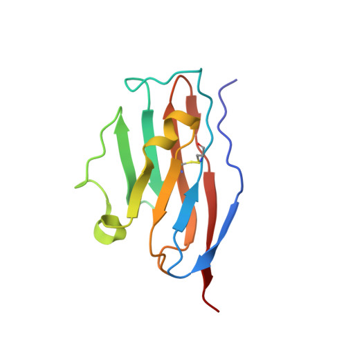
    Novel 2-arylmethoxy-4-(2,2'-dihalogen-substituted biphenyl-3-ylmethoxy) benzylamine derivatives were designed, synthesized, and evaluated in vitro and in vivo against cancers as PD-1/PD-L1 inhibitors. Through the computer-aided structural optimization and the homogeneous time-resolved fluorescence (HTRF) assay, compound A56 was found to most strongly block the PD-1/PD-L1 interaction with an IC 50 value of 2.4 ± 0.8 nM and showed the most potent activity. 1 H NMR titration results indicated that A56 can tightly bind to the PD-L1 protein with K D < 1 μM. The X-ray diffraction data for the cocrystal structure of the A56/ PD-L1 complex (3.5 Å) deciphered a novel binding mode in detail, which can account for its most potent inhibitory activity. Cell-based assays further demonstrated the strong ability of A56 as an hPD-1/hPD-L1 blocker. Especially in an hPD-L1 MC38 humanized mouse model, A56 significantly inhibited tumor growth without obvious toxicity, with a TGI rate of 55.20% (50 mg/kg, i.g.). In conclusion, A56 is a promising clinical candidate worthy of further development.


  • Organizational Affiliation
    • Lab of Chemical Biology and Molecular Drug Design, College of Pharmaceutical Science, Zhejiang University of Technology, Hangzhou 310014, China.

Macromolecules
Find similar proteins by:  (by identity cutoff)  |  3D Structure
Entity ID: 1
MoleculeChains Sequence LengthOrganismDetailsImage
Programmed cell death 1 ligand 1
A, B
116Homo sapiensMutation(s): 0 
Gene Names: CD274B7H1PDCD1L1PDCD1LG1PDL1
UniProt & NIH Common Fund Data Resources
Find proteins for Q9NZQ7 (Homo sapiens)
Explore Q9NZQ7 
Go to UniProtKB:  Q9NZQ7
PHAROS:  Q9NZQ7
GTEx:  ENSG00000120217 
Entity Groups  
Sequence Clusters30% Identity50% Identity70% Identity90% Identity95% Identity100% Identity
UniProt GroupQ9NZQ7
Sequence Annotations
Expand
  • Reference Sequence
Small Molecules
Ligands 1 Unique
IDChains Name / Formula / InChI Key2D Diagram3D Interactions
VYC (Subject of Investigation/LOI)
Query on VYC

Download Ideal Coordinates CCD File 
C [auth A]5-[[5-[[2-chloranyl-3-(2-fluorophenyl)phenyl]methoxy]-2-[(~{E})-2-hydroxyethyliminomethyl]phenoxy]methyl]pyridine-3-carbonitrile
C29 H25 Cl F N3 O3
DSELNKYPNSPLEU-UHFFFAOYSA-N
Experimental Data & Validation

Experimental Data

  • Method: X-RAY DIFFRACTION
  • Resolution: 3.50 Å
  • R-Value Free:  0.296 (Depositor), 0.300 (DCC) 
  • R-Value Work:  0.292 (Depositor), 0.290 (DCC) 
  • R-Value Observed: 0.293 (Depositor) 
Space Group: P 31 2 1
Unit Cell:
Length ( Å )Angle ( ˚ )
a = 73.459α = 90
b = 73.459β = 90
c = 95.156γ = 120
Software Package:
Software NamePurpose
REFMACrefinement
XDSdata reduction
Aimlessdata scaling
PHASERphasing

Structure Validation

View Full Validation Report


Ligand Structure Quality Assessment 


Entry History & Funding Information

Deposition Data


Funding OrganizationLocationGrant Number
National Natural Science Foundation of China (NSFC)China21877101
Foundation for Polish SciencePolandTEAM TECH CORE FACILITY/2017-4/6
National Natural Science Foundation of China (NSFC)China22177105

Revision History  (Full details and data files)

  • Version 1.0: 2023-08-02
    Type: Initial release
  • Version 1.1: 2023-08-23
    Changes: Data collection, Database references
  • Version 1.2: 2024-11-13
    Changes: Structure summary