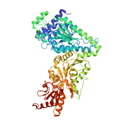
8R3P | pdb_00008r3p

Transketolase from Enterococcus faecium in complex with thiamin pyrophosphate


Experimental Data Snapshot

  • Method: X-RAY DIFFRACTION
  • Resolution: 2.90 Å
  • R-Value Free: 
    0.243 (Depositor), 0.250 (DCC) 
  • R-Value Work: 
    0.208 (Depositor), 0.210 (DCC) 
  • R-Value Observed: 
    0.209 (Depositor) 

Starting Model: in silico
View more details

wwPDB Validation   3D Report Full Report


Ligand Structure Quality Assessment 


This is version 1.1 of the entry. See complete history


Literature

Biochemical, Bioinformatic, and Structural Comparisons of Transketolases and Position of Human Transketolase in the Enzyme Evolution.

Georges, R.N.Ballut, L.Aghajari, N.Hecquet, L.Charmantray, F.Doumeche, B.

(2024) Biochemistry 63: 1460-1473

  • DOI: https://doi.org/10.1021/acs.biochem.3c00714
  • Primary Citation of Related Structures:  
    8R3O, 8R3P, 8R3Q, 8R3R, 8R3S

  • PubMed Abstract: 

    Transketolases (TKs) are key enzymes of the pentose phosphate pathway, regulating several other critical pathways in cells. Considering their metabolic importance, TKs are expected to be conserved throughout evolution. However, Tittmann et al. ( J Biol Chem , 2010 , 285(41): 31559-31570) demonstrated that Homo sapiens TK ( hs TK) possesses several structural and kinetic differences compared to bacterial TKs. Here, we study 14 TKs from pathogenic bacteria, fungi, and parasites and compare them with hs TK using biochemical, bioinformatic, and structural approaches. For this purpose, six new TK structures are solved by X-ray crystallography, including the TK of Plasmodium falciparum . All of these TKs have the same general fold as bacterial TKs. This comparative study shows that hs TK greatly differs from TKs from pathogens in terms of enzymatic activity, spatial positions of the active site, and monomer-monomer interface residues. An ubiquitous structural pattern is identified in all TKs as a six-residue histidyl crown around the TK cofactor (thiamine pyrophosphate), except for hs TK containing only five residues in the crown. Residue mapping of the monomer-monomer interface and the active site reveals that hs TK contains more unique residues than other TKs. From an evolutionary standpoint, TKs from animals (including H. sapiens ) and Schistosoma sp. belong to a distinct structural group from TKs of bacteria, plants, fungi, and parasites, mostly based on a different linker between domains, raising hypotheses regarding evolution and regulation.


  • Organizational Affiliation
    • Univ Lyon, Université Claude Bernard Lyon 1, CNRS, ICBMS UMR5246, 69622 Villeurbanne, France.

Macromolecules
Find similar proteins by:  (by identity cutoff)  |  3D Structure
Entity ID: 1
MoleculeChains Sequence LengthOrganismDetailsImage
Transketolase
A, B, C, D
680Enterococcus faeciumMutation(s): 0 
Gene Names: tktAHMPREF0351_11311
EC: 2.2.1.1
UniProt
Find proteins for A0A133CKH8 (Enterococcus faecium)
Explore A0A133CKH8 
Go to UniProtKB:  A0A133CKH8
Entity Groups  
Sequence Clusters30% Identity50% Identity70% Identity90% Identity95% Identity100% Identity
UniProt GroupA0A133CKH8
Sequence Annotations
Expand
  • Reference Sequence
Small Molecules
Ligands 2 Unique
IDChains Name / Formula / InChI Key2D Diagram3D Interactions
TPP
Query on TPP

Download Ideal Coordinates CCD File 
E [auth A],
G [auth B],
I [auth C],
K [auth D]
THIAMINE DIPHOSPHATE
C12 H19 N4 O7 P2 S
AYEKOFBPNLCAJY-UHFFFAOYSA-O
MG
Query on MG

Download Ideal Coordinates CCD File 
F [auth A],
H [auth B],
J [auth C],
L [auth D]
MAGNESIUM ION
Mg
JLVVSXFLKOJNIY-UHFFFAOYSA-N
Experimental Data & Validation

Experimental Data

  • Method: X-RAY DIFFRACTION
  • Resolution: 2.90 Å
  • R-Value Free:  0.243 (Depositor), 0.250 (DCC) 
  • R-Value Work:  0.208 (Depositor), 0.210 (DCC) 
  • R-Value Observed: 0.209 (Depositor) 
Space Group: C 1 2 1
Unit Cell:
Length ( Å )Angle ( ˚ )
a = 249.66α = 90
b = 68.22β = 110.71
c = 165.46γ = 90
Software Package:
Software NamePurpose
PHENIXrefinement
PHENIXrefinement
XDSdata reduction
XSCALEdata scaling
PHENIXphasing

Structure Validation

View Full Validation Report


Ligand Structure Quality Assessment 


Entry History & Funding Information

Deposition Data


Funding OrganizationLocationGrant Number
Other government--

Revision History  (Full details and data files)

  • Version 1.0: 2024-05-22
    Type: Initial release
  • Version 1.1: 2024-12-04
    Changes: Database references, Structure summary