8RAQ | pdb_00008raq

Crystal structure of Mycobacterium tuberculosis MmaA1 with S-adenosyl methionine (SAM)


Experimental Data Snapshot

  • Method: X-RAY DIFFRACTION
  • Resolution: 1.40 Å
  • R-Value Free: 
    0.171 (Depositor), 0.173 (DCC) 
  • R-Value Work: 
    0.150 (Depositor), 0.152 (DCC) 
  • R-Value Observed: 
    0.150 (Depositor) 

Starting Model: in silico
View more details

wwPDB Validation   3D Report Full Report


Ligand Structure Quality Assessment 


This is version 1.2 of the entry. See complete history


Literature

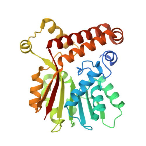
Crystal structures of the mycolic acid methyl transferase 1 (MmaA1) from Mycobacterium tuberculosis in the apo-form and in complex with different cofactors reveal unique features for substrate binding.

Chaudhary, B.Kobakhidze, G.Wachelder, L.Mazumdar, P.A.Dong, G.Madhurantakam, C.

(2025) J Biomol Struct Dyn : 1-10

  • DOI: https://doi.org/10.1080/07391102.2025.2483952
  • Primary Citation of Related Structures:  
    8RAQ, 8RBD, 8RBE, 8RBL

  • PubMed Abstract: 

    Mycolic acid methyl transferase 1 (MmaA1) protein from Mycobacterium tuberculosis plays a crucial role in the biosynthesis of cell wall mycolic acids that aid in survival of the bacteria under adverse conditions. The enzyme converts a cis to a trans olefin and adds a methyl group at the proximal position of both methoxy and keto-mycolic acid chains. Here we report the crystal structures of apo-MmaA1 and complexes with the cofactor S-adenosylmethionine (SAM), the end-product of methylation reactions - S-adenosylhomocysteine (SAH), and the nucleoside analog Sinefungin (SFG) at 1.4-1.9 Å resolution. These structures reveal the typical seven-stranded α/β fold accompanied by other α-helical embellishments. A dynamic labile loop across the cofactor binding site in the apo-form became relatively rigid upon binding of SAM or SFG but remained labile in the SAH-bound form. A comprehensive analysis of the binding pattern of SAM with MmaA1 reveals critical residues involved in the hydrogen bond interactions with the cofactor, most of which are conserved across other methyltransferases. We also observed a highly conserved cysteine residue (C268) packed against the inner part of the substrate entry channel. C268 is in the reduced state in the SAM-bound but oxidized in the SAH-bound structure. The bulkier sidechain of the oxidized C268 significantly blocks the substrate-binding channel, which might serve as a regulator to control substrate binding and/or selectivity. This atomic view of this critical methyltransferase will build a basis for the identification of small molecule inhibitors against M. tuberculosis.


  • Organizational Affiliation
    • Structural and Molecular Biology Laboratory (SMBL), Department of Biotechnology, TERI School of Advanced Studies (TERI SAS), New Delhi, India.

Macromolecules
Find similar proteins by:  (by identity cutoff)  |  3D Structure
Entity ID: 1
MoleculeChains Sequence LengthOrganismDetailsImage
Mycolic acid methyltransferase MmaA1A,
B [auth A0A0]
287Mycobacterium tuberculosis H37RvMutation(s): 0 
Gene Names: mmaA1
EC: 2.1.1
UniProt
Find proteins for P9WPB1 (Mycobacterium tuberculosis (strain ATCC 25618 / H37Rv))
Explore P9WPB1 
Go to UniProtKB:  P9WPB1
Entity Groups  
Sequence Clusters30% Identity50% Identity70% Identity90% Identity95% Identity100% Identity
UniProt GroupP9WPB1
Sequence Annotations
Expand
  • Reference Sequence
Small Molecules
Ligands 4 Unique
IDChains Name / Formula / InChI Key2D Diagram3D Interactions
SAM (Subject of Investigation/LOI)
Query on SAM

Download Ideal Coordinates CCD File 
C [auth A],
I [auth A0A0]
S-ADENOSYLMETHIONINE
C15 H22 N6 O5 S
MEFKEPWMEQBLKI-FCKMPRQPSA-N
GOL
Query on GOL

Download Ideal Coordinates CCD File 
D [auth A],
J [auth A0A0]
GLYCEROL
C3 H8 O3
PEDCQBHIVMGVHV-UHFFFAOYSA-N
EDO
Query on EDO

Download Ideal Coordinates CCD File 
E [auth A],
F [auth A],
H [auth A0A0],
K [auth A0A0]
1,2-ETHANEDIOL
C2 H6 O2
LYCAIKOWRPUZTN-UHFFFAOYSA-N
NA
Query on NA

Download Ideal Coordinates CCD File 
G [auth A]SODIUM ION
Na
FKNQFGJONOIPTF-UHFFFAOYSA-N
Experimental Data & Validation

Experimental Data

  • Method: X-RAY DIFFRACTION
  • Resolution: 1.40 Å
  • R-Value Free:  0.171 (Depositor), 0.173 (DCC) 
  • R-Value Work:  0.150 (Depositor), 0.152 (DCC) 
  • R-Value Observed: 0.150 (Depositor) 
Space Group: P 1 21 1
Unit Cell:
Length ( Å )Angle ( ˚ )
a = 41.384α = 90
b = 74.734β = 90.74
c = 96.773γ = 90
Software Package:
Software NamePurpose
MxCuBEdata collection
PHENIXrefinement
XDSdata reduction
XSCALEdata scaling
PHENIXphasing

Structure Validation

View Full Validation Report


Ligand Structure Quality Assessment 


Entry History & Funding Information

Deposition Data


Funding OrganizationLocationGrant Number
Austrian Science FundAustriaI5960-B2

Revision History  (Full details and data files)

  • Version 1.0: 2024-12-11
    Type: Initial release
  • Version 1.1: 2025-05-07
    Changes: Database references, Structure summary
  • Version 1.2: 2025-06-04
    Changes: Database references, Structure summary