8RTJ | pdb_00008rtj

X-ray structure of lysozyme obtained upon reaction with [VIVO(8-HQ)2] in ethylene glycol


Experimental Data Snapshot

  • Method: X-RAY DIFFRACTION
  • Resolution: 1.27 Å
  • R-Value Free: 
    0.197 (Depositor), 0.202 (DCC) 
  • R-Value Work: 
    0.174 (Depositor), 0.180 (DCC) 

Starting Model: experimental
View more details

wwPDB Validation   3D Report Full Report


Ligand Structure Quality Assessment 


This is version 1.1 of the entry. See complete history


Literature

Protein-Protein Stabilization in V IV O/8-Hydroxyquinoline-Lysozyme Adducts.

Paolillo, M.Ferraro, G.Pisanu, F.Marechal, J.D.Sciortino, G.Garribba, E.Merlino, A.

(2024) Chemistry 30: e202401712-e202401712

  • DOI: https://doi.org/10.1002/chem.202401712
  • Primary Citation of Related Structures:  
    8RTJ, 8RTK

  • PubMed Abstract: 

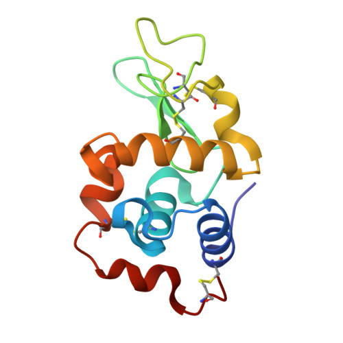
    The binding of the potential drug [V IV O(8-HQ) 2 ], where 8-HQ is 8-hydroxyquinolinato, with hen egg white lysozyme (HEWL) was evaluated through spectroscopic (electron paramagnetic resonance, EPR, and UV-visible), spectrometric (electrospray ionization-mass spectrometry, ESI-MS), crystallographic (X-ray diffraction, XRD), and computational (DFT and docking) studies. ESI-MS indicates the interaction of [V IV O(8-HQ)(H 2 O)] + and [V IV O(8-HQ) 2 (H 2 O)] species with HEWL. Room temperature EPR spectra suggest both covalent and non-covalent binding of the two different V-containing fragments. XRD analyses confirm these findings, showing that [V IV O(8-HQ)(H 2 O)] + interacts covalently with the solvent exposed Asp119, while cis-[V IV O(8-HQ) 2 (H 2 O)] non-covalently with Arg128 and Lys96 from a symmetry mate. The covalent binding of [V IV O(8-HQ)(H 2 O)] + to Asp119 is favored by a π-π contact with Trp62 and a H-bond with Asn103 of a symmetry-related molecule. Additionally, the covalent binding of V V O 2 + to Asp48 and non-covalent binding of other V-containing fragments to Arg5, Cys6, and Glu7 are revealed. Molecular docking indicates that, in the absence of the interactions occurring at the protein-protein interface close to Asp119, the covalent binding to Glu35 or Asp52 should be preferred. Such a protein-protein stabilization could be more common than what believed up today, at least in the solid state, and should be considered in the characterization of metal-protein adducts.


  • Organizational Affiliation
    • Department of Chemical Sciences, University of Naples 'Federico II', Complesso Universitario di Monte Sant'Angelo, Via Cintia, I-80126, Napoli, Italy.

Macromolecules
Find similar proteins by:  (by identity cutoff)  |  3D Structure
Entity ID: 1
MoleculeChains Sequence LengthOrganismDetailsImage
Lysozyme CA [auth AAA]129Gallus gallusMutation(s): 0 
EC: 3.2.1.17
UniProt
Find proteins for P00698 (Gallus gallus)
Explore P00698 
Go to UniProtKB:  P00698
Entity Groups  
Sequence Clusters30% Identity50% Identity70% Identity90% Identity95% Identity100% Identity
UniProt GroupP00698
Sequence Annotations
Expand
  • Reference Sequence
Small Molecules
Ligands 4 Unique
IDChains Name / Formula / InChI Key2D Diagram3D Interactions
I3Y (Subject of Investigation/LOI)
Query on I3Y

Download Ideal Coordinates CCD File 
C [auth AAA]2,2-bis($l^{1}-oxidanyl)-3-oxa-1$l^{4}-aza-2$l^{4}-vanadatricyclo[6.3.1.0^{4,12}]dodeca-1(12),4,6,8,10-pentaene
C9 H8 N O3 V
VHSPINZPWRCWRI-UHFFFAOYSA-K
NO3
Query on NO3

Download Ideal Coordinates CCD File 
E [auth AAA]
F [auth AAA]
G [auth AAA]
H [auth AAA]
I [auth AAA]
E [auth AAA],
F [auth AAA],
G [auth AAA],
H [auth AAA],
I [auth AAA],
J [auth AAA],
K [auth AAA],
L [auth AAA],
M [auth AAA]
NITRATE ION
N O3
NHNBFGGVMKEFGY-UHFFFAOYSA-N
V
Query on V

Download Ideal Coordinates CCD File 
D [auth AAA]VANADIUM ION
V
KOKKJWHERHSKEB-UHFFFAOYSA-N
NA
Query on NA

Download Ideal Coordinates CCD File 
B [auth AAA]SODIUM ION
Na
FKNQFGJONOIPTF-UHFFFAOYSA-N
Experimental Data & Validation

Experimental Data

  • Method: X-RAY DIFFRACTION
  • Resolution: 1.27 Å
  • R-Value Free:  0.197 (Depositor), 0.202 (DCC) 
  • R-Value Work:  0.174 (Depositor), 0.180 (DCC) 
Space Group: P 43 21 2
Unit Cell:
Length ( Å )Angle ( ˚ )
a = 78.14α = 90
b = 78.14β = 90
c = 37.52γ = 90
Software Package:
Software NamePurpose
REFMACrefinement
autoPROCdata reduction
autoPROCdata scaling
PHASERphasing

Structure Validation

View Full Validation Report


Ligand Structure Quality Assessment 


Entry History & Funding Information

Deposition Data


Funding OrganizationLocationGrant Number
University of Naples Federico IIItaly--

Revision History  (Full details and data files)

  • Version 1.0: 2025-02-12
    Type: Initial release
  • Version 1.1: 2025-07-16
    Changes: Database references