


Atomic structure of truncated worm GdH
Mourao, A., Sattler, M., Geerlof, A.To be published.
Experimental Data Snapshot
Starting Model: experimental
View more details
wwPDB Validation 3D Report Full Report

Entity ID: 1 | |||||
|---|---|---|---|---|---|
| Molecule | Chains | Sequence Length | Organism | Details | Image |
| Glutamate dehydrogenase | 537 | Heligmosomoides polygyrus | Mutation(s): 0 Gene Names: HPBE_LOCUS9300 | 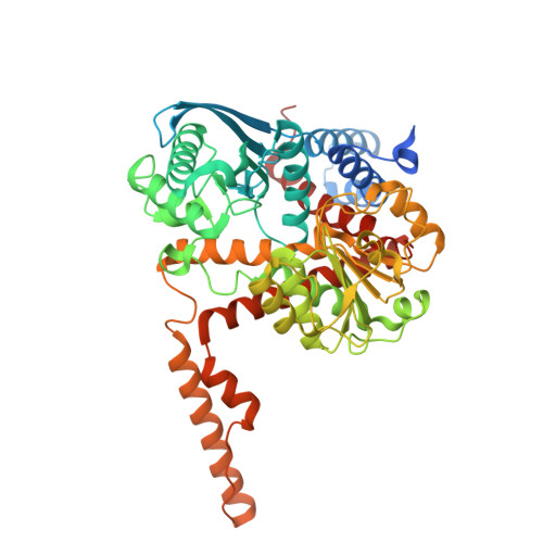 | |
UniProt | |||||
Find proteins for A0A183FP08 (Heligmosomoides polygyrus) Explore A0A183FP08 Go to UniProtKB: A0A183FP08 | |||||
Entity Groups | |||||
| Sequence Clusters | 30% Identity50% Identity70% Identity90% Identity95% Identity100% Identity | ||||
| UniProt Group | A0A183FP08 | ||||
Sequence AnnotationsExpand | |||||
| |||||
| Length ( Å ) | Angle ( ˚ ) |
|---|---|
| a = 151.245 | α = 90 |
| b = 151.245 | β = 90 |
| c = 146.043 | γ = 120 |
| Software Name | Purpose |
|---|---|
| PHENIX | refinement |
| XDS | data reduction |
| Aimless | data scaling |
| PHASER | phasing |
| Funding Organization | Location | Grant Number |
|---|---|---|
| Helmholtz Association | Germany | -- |


