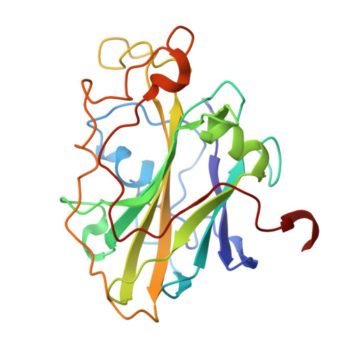
Anions and citrate inhibit LsAA9A, a lytic polysaccharide monooxygenase (LPMO).
Di Domenico, V., Theibich, Y., Brander, S., Berrin, J.G., Johansen, K.S., Frandsen, K.E.H., Lo Leggio, L.(2025) FEBS J 292: 4375-4389
- PubMed: 40424050
- DOI: https://doi.org/10.1111/febs.70138
- Primary Citation of Related Structures:
8S3F, 8S3L, 9EQE - PubMed Abstract:
Lytic polysaccharide monooxygenases (LPMOs) are oxidative enzymes that break the glycosidic linkage in recalcitrant polysaccharides such as cellulose and chitin. The LPMO LsAA9A (AA9 family lytic polysaccharide monooxygenase A) from the basidiomycete fungus Lentinus similis is biochemically and structurally well characterized, with crystallographic complexes with oligosaccharides having been obtained. Chloride ions from the crystallization solution are known to bind to the LsAA9A-substrate complex in crystals at the copper equatorial coordinating position, where activation of the co-substrate oxygen species is expected. An investigation of the effect of high concentration salts on LsAA9A activity showed that salts containing chloride and other halide anions, except for fluoride, had a clear inhibitory effect on the activity at concentrations > 100 mm, although chloride ions are known to increase the LPMO affinity for oligosaccharide binding. Surprisingly, LsAA9A crystals can be transferred for short times to considerably different chemical environments, allowing crystallographic analysis at reduced chloride concentrations. Unfortunately, these washing steps do not eliminate the chloride binding at the copper equatorial coordinating position. Furthermore, we observed that citrate buffer, also present, bound under these changed chemical conditions at the copper active site. This interaction completely blocks access to the oligosaccharide substrate and is additionally supported here by citrate inhibition of LsAA9A activities against azurine cross-linked hydroxyethylcellulose (AZCL-HEC), tamarind xyloglucan, and cellopentaose. The conclusions from our study indicate that citrate should be absolutely avoided in LPMO research, not only because of possible abstraction of copper ions from the LPMO active site but also because it might directly compete with binding of LPMOs to their target substrates.
- Department of Chemistry, University of Copenhagen, Denmark.
Organizational Affiliation: