8S4Y | pdb_00008s4y

Crystal structure of an APP-talin (F2F3) chimera


Experimental Data Snapshot

  • Method: X-RAY DIFFRACTION
  • Resolution: 2.70 Å
  • R-Value Free: 
    0.267 (Depositor), 0.268 (DCC) 
  • R-Value Work: 
    0.211 (Depositor), 0.218 (DCC) 
  • R-Value Observed: 
    0.217 (Depositor) 

Starting Model: experimental
View more details

wwPDB Validation   3D Report Full Report


This is version 1.1 of the entry. See complete history


Literature

The structure of an amyloid precursor protein/talin complex indicates a mechanical basis of Alzheimer's disease.

Ellis, C.Ward, N.L.Rice, M.Ball, N.J.Walle, P.Najdek, C.Kilinc, D.Lambert, J.C.Chapuis, J.Goult, B.T.

(2024) Open Biol 14: 240185-240185

  • DOI: https://doi.org/10.1098/rsob.240185
  • Primary Citation of Related Structures:  
    8S4Y

  • PubMed Abstract: 

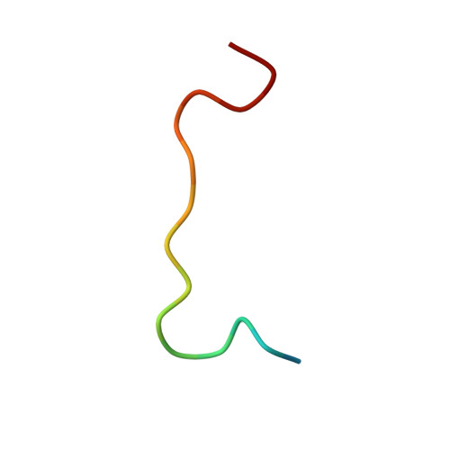
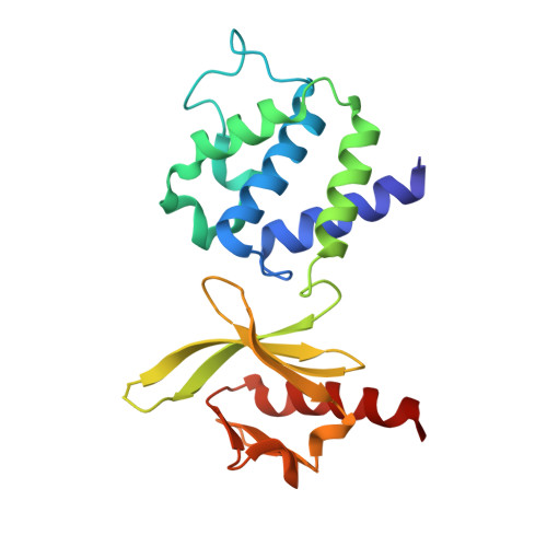
    Misprocessing of amyloid precursor protein (APP) is one of the major causes of Alzheimer's disease. APP comprises a large extracellular region, a single transmembrane helix and a short cytoplasmic tail containing an NPxY motif (normally referred to as the YENPTY motif). Talins are synaptic scaffold proteins that connect the cytoskeletal machinery to the plasma membrane via binding NPxY motifs in the cytoplasmic tail of integrins. Here, we report the crystal structure of an APP/talin1 complex identifying a new way to couple the cytoskeletal machinery to synaptic sites through APP. Proximity ligation assay (PLA) confirmed the close proximity of talin1 and APP in primary neurons, and talin1 depletion had a dramatic effect on APP processing in cells. Structural modelling reveals APP might form an extracellular meshwork that mechanically couples the cytoskeletons of the pre- and post-synaptic compartments. We propose APP processing represents a mechanical signalling pathway whereby under tension, the cleavage sites in APP have varying accessibility to cleavage by secretases. This leads us to propose a new hypothesis for Alzheimer's, where misregulated APP dynamics result in loss of the mechanical integrity of the synapse, corruption and loss of mechanical binary data, and excessive generation of toxic plaque-forming Aβ42 peptide.


  • Organizational Affiliation
    • School of Biosciences, University of Kent, Canterbury, Kent CT2 7NJ, UK.

Macromolecules

Find similar proteins by:  Sequence   |   3D Structure  

Entity ID: 1
MoleculeChains Sequence LengthOrganismDetailsImage
C3117Mus musculusMutation(s): 0 
Gene Names: AppA4AD1
UniProt & NIH Common Fund Data Resources
Find proteins for P12023 (Mus musculus)
Explore P12023 
Go to UniProtKB:  P12023
IMPC:  MGI:88059
Entity Groups  
Sequence Clusters30% Identity50% Identity70% Identity90% Identity95% Identity100% Identity
UniProt GroupP12023
Sequence Annotations
Expand
  • Reference Sequence
Find similar proteins by:  (by identity cutoff)  |  3D Structure
Entity ID: 2
MoleculeChains Sequence LengthOrganismDetailsImage
Talin-1192Mus musculusMutation(s): 0 
Gene Names: TLN1TLN
UniProt
Find proteins for P54939 (Gallus gallus)
Explore P54939 
Go to UniProtKB:  P54939
Entity Groups  
Sequence Clusters30% Identity50% Identity70% Identity90% Identity95% Identity100% Identity
UniProt GroupP54939
Sequence Annotations
Expand
  • Reference Sequence
Experimental Data & Validation

Experimental Data

  • Method: X-RAY DIFFRACTION
  • Resolution: 2.70 Å
  • R-Value Free:  0.267 (Depositor), 0.268 (DCC) 
  • R-Value Work:  0.211 (Depositor), 0.218 (DCC) 
  • R-Value Observed: 0.217 (Depositor) 
Space Group: P 21 21 21
Unit Cell:
Length ( Å )Angle ( ˚ )
a = 60.388α = 90
b = 64.081β = 90
c = 65.191γ = 90
Software Package:
Software NamePurpose
PHENIXrefinement
DIALSdata scaling
xia2data reduction
PHASERphasing

Structure Validation

View Full Validation Report


Entry History & Funding Information

Deposition Data


Funding OrganizationLocationGrant Number
Cancer Research UKUnited Kingdom--

Revision History  (Full details and data files)

  • Version 1.0: 2024-10-16
    Type: Initial release
  • Version 1.1: 2025-04-30
    Changes: Database references