A Structural Investigation of the Interaction between a GC-376-Based Peptidomimetic PROTAC and Its Precursor with the Viral Main Protease of Coxsackievirus B3.
De Santis, A., Grifagni, D., Orsetti, A., Lenci, E., Rosato, A., D'Onofrio, M., Trabocchi, A., Ciofi-Baffoni, S., Cantini, F., Calderone, V.(2024) Biomolecules 14
- PubMed: 39456193
- DOI: https://doi.org/10.3390/biom14101260
- Primary Citation of Related Structures:
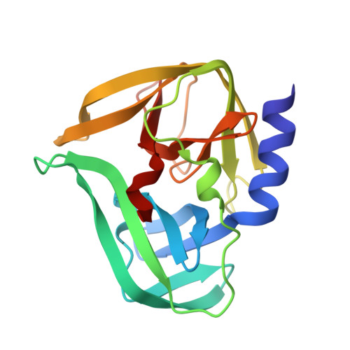
8S6F - PubMed Abstract:
The conservation of the main protease in viral genomes, combined with the absence of a homologous protease in humans, makes this enzyme family an ideal target for developing broad-spectrum antiviral drugs with minimized host toxicity. GC-376, a peptidomimetic 3CL protease inhibitor, has shown significant efficacy against coronaviruses. Recently, a GC-376-based PROTAC was developed to target and induce the proteasome-mediated degradation of the dimeric SARS-CoV-2 3CL Pro protein. Extending this approach, the current study investigates the application of the GC-376 PROTAC to the 3C Pro protease of enteroviruses, specifically characterizing its interaction with CVB3 3C Pro through X-ray crystallography, NMR (Nuclear Magnetic Resonance) and biochemical techniques. The crystal structure of CVB3 3C Pro bound to the GC-376 PROTAC precursor was obtained at 1.9 Å resolution. The crystallographic data show that there are some changes between the binding of CVB3 3C Pro and SARS-CoV-2 3CL Pro , but the overall similarity is strong (RMSD on C-alpha 0.3 Å). The most notable variation is the orientation of the benzyloxycarbonyl group of GC-376 with the S4 subsite of the proteases. NMR backbone assignment of CVB3 3C Pro bound and unbound to the GC-376 PROTAC precursor (80% and 97%, respectively) was obtained. This information complemented the investigation, by NMR, of the interaction of CVB3 3C Pro with the GC-376 PROTAC, and its precursor allows us to define that the GC-376 PROTAC binds to CVB3 3C Pro in a mode very similar to that of the precursor. The NMR relaxation data indicate that a quench of dynamics of a large part of the protein backbone involving the substrate-binding site and surrounding regions occurs upon GC-376 PROTAC precursor binding. This suggests that the substrate cavity, by sampling different backbone conformations in the absence of the substrate, is able to select the suitable one necessary to covalently bind the substrate, this being the latter reaction, which is the fundamental step required to functionally activate the enzymatic reaction. The inhibition activity assay showed inhibition potency in the micromolar range for GC-376 PROTAC and its precursor. Overall, we can conclude that the GC-376 PROTAC fits well within the binding sites of both proteases, demonstrating its potential as a broad-spectrum antiviral agent.
- Magnetic Resonance Center CERM, University of Florence, Via Luigi Sacconi 6, Sesto Fiorentino, 50019 Florence, Italy.
Organizational Affiliation: