8S83 | pdb_00008s83

Domains 3 and 6 of Sap S-layer protein from Bacillus anthracis


Experimental Data Snapshot

  • Method: X-RAY DIFFRACTION
  • Resolution: 2.07 Å
  • R-Value Free: 
    0.258 (Depositor), 0.257 (DCC) 
  • R-Value Work: 
    0.201 (Depositor), 0.202 (DCC) 
  • R-Value Observed: 
    0.204 (Depositor) 

Starting Model: experimental
View more details

wwPDB Validation   3D Report Full Report


This is version 1.1 of the entry. See complete history


Literature

Architecture of the Sap S-layer of Bacillus anthracis revealed by integrative structural biology.

Sogues, A.Leigh, K.Halingstad, E.V.Van der Verren, S.E.Cecil, A.J.Fioravanti, A.Pak, A.J.Kudryashev, M.Remaut, H.

(2024) Proc Natl Acad Sci U S A 121: e2415351121-e2415351121

  • DOI: https://doi.org/10.1073/pnas.2415351121
  • Primary Citation of Related Structures:  
    8RX2, 8S80, 8S83, 9G93

  • PubMed Abstract: 

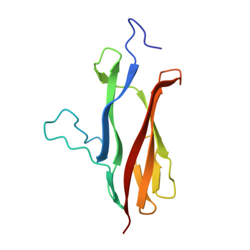
    Bacillus anthracis is a spore-forming gram-positive bacterium responsible for anthrax, an infectious disease with a high mortality rate and a target of concern due to bioterrorism and long-term site contamination. The entire surface of vegetative cells in exponential or stationary growth phase is covered in proteinaceous arrays called S-layers, composed of Sap or EA1 protein, respectively. The Sap S-layer represents an important virulence factor and cell envelope support structure whose paracrystalline nature is essential for its function. However, the spatial organization of Sap in its lattice state remains elusive. Here, we employed cryoelectron tomography and subtomogram averaging to obtain a map of the Sap S-layer from tubular polymers that revealed a conformational switch between the postassembly protomers and the previously available X-ray structure of the condensed monomers. To build and validate an atomic model of the lattice within this map, we used a combination of molecular dynamics simulations, X-ray crystallography, cross-linking mass spectrometry, and biophysics in an integrative structural biology approach. The Sap lattice model produced recapitulates a close-to-physiological arrangement, reveals high-resolution details of lattice contacts, and sheds light on the mechanisms underlying the stability of the Sap layer.


  • Organizational Affiliation
    • Structural and Molecular Microbiology, Vlaams Instituut voor Biotechnologie (VIB)-Vrije Universiteit Brussel (VUB) Center for Structural Biology, Vlaams Instituut voor Biotechnologie, Brussels 1050, Belgium.

Macromolecules
Find similar proteins by:  (by identity cutoff)  |  3D Structure
Entity ID: 1
MoleculeChains Sequence LengthOrganismDetailsImage
S-layer protein sap
A, B
113Bacillus anthracisMutation(s): 0 
Gene Names: sapBA_0885GBAA_0885BAS0841
UniProt
Find proteins for P49051 (Bacillus anthracis)
Explore P49051 
Go to UniProtKB:  P49051
Entity Groups  
Sequence Clusters30% Identity50% Identity70% Identity90% Identity95% Identity100% Identity
UniProt GroupP49051
Sequence Annotations
Expand
  • Reference Sequence
Find similar proteins by:  (by identity cutoff)  |  3D Structure
Entity ID: 2
MoleculeChains Sequence LengthOrganismDetailsImage
S-layer protein sap
C, D
109Bacillus anthracisMutation(s): 0 
Gene Names: sapBA_0885GBAA_0885BAS0841
UniProt
Find proteins for P49051 (Bacillus anthracis)
Explore P49051 
Go to UniProtKB:  P49051
Entity Groups  
Sequence Clusters30% Identity50% Identity70% Identity90% Identity95% Identity100% Identity
UniProt GroupP49051
Sequence Annotations
Expand
  • Reference Sequence
Experimental Data & Validation

Experimental Data

  • Method: X-RAY DIFFRACTION
  • Resolution: 2.07 Å
  • R-Value Free:  0.258 (Depositor), 0.257 (DCC) 
  • R-Value Work:  0.201 (Depositor), 0.202 (DCC) 
  • R-Value Observed: 0.204 (Depositor) 
Space Group: P 1 21 1
Unit Cell:
Length ( Å )Angle ( ˚ )
a = 40.154α = 90
b = 61.01β = 91.5
c = 97.658γ = 90
Software Package:
Software NamePurpose
PHENIXrefinement
XDSdata reduction
Aimlessdata scaling
PHASERphasing

Structure Validation

View Full Validation Report


Entry History & Funding Information

Deposition Data


Funding OrganizationLocationGrant Number
European Molecular Biology Organization (EMBO)European UnionALTF 709-2021

Revision History  (Full details and data files)

  • Version 1.0: 2024-11-20
    Type: Initial release
  • Version 1.1: 2024-12-18
    Changes: Database references